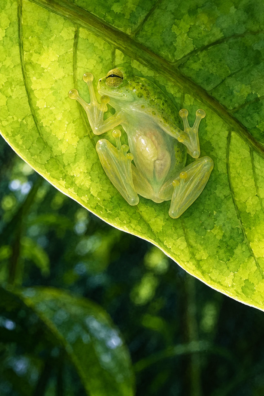
A sleeping glassfrog on a leaf is not simply painted to match the leaf. Part of the background can pass visually through it. Even more surprisingly, its transparency changes with its state: the quiet daytime animal can become harder to see than the active frog. To explain that change, researchers had to follow something normally expected to remain conspicuous—red blood cells.
Glassfrogs have translucent tissues, especially visible from below, but blood poses a problem for a transparent vertebrate. Red blood cells contain hemoglobin, which interacts strongly with light. A body full of circulating red cells is difficult to make optically unobtrusive merely by removing pigment from the skin. Transparency is therefore a whole-body problem, not just a skin color.
The blood was hidden, not removed from existence
A 2022 study in Science investigated Fleischmann's glassfrog, Hyalinobatrachium fleischmanni. In the resting state, frogs sequestered a large fraction of their red blood cells in the liver. When active, more cells returned to circulation. This reversible redistribution helps explain how a frog can retain the blood cells needed for activity while reducing their optical effect during rest.
The American Museum of Natural History's account reports that resting frogs increased transparency roughly two- to threefold while removing nearly 90 percent of red cells from circulation. Those numbers belong to the studied animals and measurements; they are not a guarantee for every frog called a glassfrog, every tissue, or every lighting condition. The liver's reflective covering helps conceal the concentrated cells.
This is a rearrangement of visibility. Concentrating a strongly light-interacting material in a concealed compartment can reduce how widely it reveals the body. Think of the difference between spreading dark beads through a clear container and gathering them behind an opaque patch. The analogy explains the optical logic, although it does not explain how a living frog regulates circulation or maintains its tissues.
How do you find blood without making the frog stop being transparent?
An ordinary photograph records appearance, but it cannot directly establish where the blood went. Researchers used photoacoustic imaging among their methods. Absorbed light produces a small acoustic signal that can be detected and used to localize absorbers such as hemoglobin. The Duke research summary connects the imaging work with the biological discovery.
The measurement problem matters because the state being measured is fragile. Handling, anesthesia, or activity could change a physiological condition. If the interesting phenomenon belongs to a resting animal, an invasive measurement that changes rest could obscure it. A good technique must therefore do more than produce a sharp image; it must preserve enough of the relevant state for the image to answer the question.
The study also raises unresolved physiology. Packing so many blood cells together without the usual harmful consequences is remarkable. But observing the natural outcome does not by itself identify every mechanism that prevents problems, and it does not establish a treatment for human blood clotting. A useful research question can be motivated by the frog without assuming that its solution transfers directly to another animal.
Transparency is not the same as invisibility
An earlier 2020 study of glassfrog camouflage examined imperfect transparency and a proposed effect called edge diffusion. Translucent legs can take on the brightness of the background more readily than the body, reducing the abruptness of the animal's outline. That is different from the later discovery of changing red-cell distribution. One concerns how partial transparency can conceal shape; the other identifies a physiological way of changing transparency.
A transparent animal can still reflect light, cast a shadow, distort a background, or retain opaque organs. For a predator, the relevant question is not whether the frog has vanished from physics. It is whether the visual information is sufficient to detect and recognize prey under real viewing conditions.
The generated image here is similarly limited. It evokes a frog beneath a leaf and the delicacy of translucent limbs. Its internal hints are artistic interpretation. For the actual distribution of blood cells, follow the paper's measurements and figures rather than reading anatomy from generated artwork.
Once those distinctions are clear, the sleeping frog becomes more impressive. It is not a passive green ornament that happens to be see-through. Its apparent stillness includes a substantial reversible physiological change. Resting on the same kind of leaf, an animal can alter the way its interior contributes to the view from outside. The camouflage is partly a matter of where the body puts its blood.
Follow the evidence: Taboada and colleagues: blood concealment in glassfrogs; American Museum of Natural History: transparent camouflage; Duke: discovering the transparency mechanism; Barnett and colleagues: imperfect transparency and edge diffusion.
More curious connections, art and independent projects at Incoprea.