Showing posts with label Cell Biology. Show all posts

Sunday, October 25, 2026

The Microscopic Thread That Turns Inside Out to Sting

A sea anemone's tentacle can look soft and uneventful. At its surface, however, specialized cells contain compact structures ready for an extraordinary transformation. A thread that was packed inside a capsule can discharge and turn inside out.

“Tiny harpoon” captures the suddenness, but misses much of the mechanism. The apparatus changes shape as it operates. Its unfolding architecture is part of how it works.

Small translucent hydroid polyp with tentacles extended from a seagrass blade.
Cnidarian tentacles carry specialized cells whose microscopic capsules can discharge an everting thread.

A cell containing a single-use capsule

Cnidarians include anemones, corals, jellyfish, and hydroids. Their specialized cnidocytes contain different kinds of capsules. The stinging capsules called nematocysts are one part of this diverse equipment.

The capsule and the cell are different levels of organization. A nematocyst is an organelle within a cell, rather than a free-living creature. Its stored thread and capsule must first be built by biological processes before discharge becomes possible.

The Stowers Institute's account of its researchers' work describes a system combining osmotic pressure with elastic energy stored in several substructures. Pressure supplies a powerful start, while the organization of the packed apparatus contributes to its subsequent transformation.

That arrangement separates preparation from action. A cell invests in making and loading the structure. When a suitable trigger arrives, it can release stored energy much faster than it could construct a new apparatus on demand.

Three phases, rather than one flying object

A 2022 study of the anemone Nematostella vectensis combined imaging methods to reconstruct the organization of a particular nematocyst type. Its researchers identified three main phases: shaft discharge, shaft eversion, and tubule eversion.

First, a tightly packed shaft is expelled. Next, its filaments uncoil and turn inside out, producing an elongated passage. The thinner tubule then everts through that passage. In this nematocyst, the shaft is both an initial penetrating structure and a protective route for the more delicate tubule.

The authors used three-dimensional electron microscopy alongside recordings of discharge. Their reconstruction links what was packed inside to what appeared outside. It is a mechanism for the studied structure, rather than an assertion that every cnidarian capsule has identical internal parts.

That specificity is valuable. Different nematocysts can perform different tasks, and comparing their architectures helps explain why a single familiar metaphor cannot stand in for the entire group.

Which part was extraordinarily fast?

The speed deserves careful reading. In 2006 research on Hydra stenoteles, specialized high-speed imaging resolved an initial discharge phase on a timescale as short as 700 nanoseconds. That is less than a millionth of a second.

The number describes an exceptionally rapid stage in the studied system. It should not be attached to the complete sequence in every anemone or jellyfish. The 2022 work explicitly distinguishes the faster Hydra mechanism from the slower shaft eversion it observed in Nematostella.

Whenever a biological speed sounds unbelievable, ask what began the stopwatch and what stopped it. Initial acceleration, full extension, and the animal's entire response are different measurements. The distinction preserves the achievement rather than diminishing it.

Why it does not fire at every disturbance

A one-use capsule needs more than an efficient launch. It also needs appropriate control.

A 2020 study of Nematostella investigated a specialized voltage-gated calcium channel in its stinging cells. The channel's properties suppress responses to irrelevant mechanical disturbances. Prey-related chemical signals can relieve that inhibition, allowing a mechanical stimulus to help trigger discharge.

This is sensory filtering at the cellular level. The animal lives in moving water; movement alone would be an expensive reason to expend every capsule. Integrating cues helps connect the available apparatus to a biologically relevant encounter.

Even that control is not one universal rule. Later research comparing Nematostella with Exaiptasia diaphana found differences in channel behavior and stinging responses. The comparison associated more tightly filtered predatory responses with one system and more readily activated defensive responses with the other.

The full story therefore has two intertwined achievements. A cell prepares a structure capable of rapid mechanical change, and sensory machinery helps determine when that structure is used. The quiet tentacle holds both a loaded geometry and a decision about contact with the world.

Read the research

Keep exploring

Discover more art, ideas, and curious things at Incoprea.

Thursday, October 22, 2026

Planarians Need Instructions as Well as Stem Cells to Rebuild

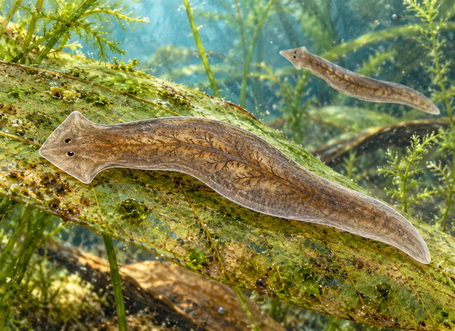
A magnified intact flatworm with a broad triangular head glides across an underwater leaf.
Conceptual natural-history illustration; internal shading is interpretive, not microscopy or gene-expression data.

A regenerating animal has more to do than make new cells. It must make the right kinds of cells, put them in appropriate places, and connect them into a body that works. A freshwater planarian makes that problem unusually visible. Its capacity to replace missing tissue forces a question that a simple “it grows back” leaves unanswered: how does the new tissue know where it belongs?

The word “know” is shorthand. No cell consults a miniature picture of the completed worm. Researchers investigate signals, gene activity, cell populations, and responses to injury. The aim is to explain how local biological processes produce an organized result.

Material and instructions are different requirements

The Reddien Lab's research overview separates two central systems. Planarian neoblasts are proliferating cells that include pluripotent stem cells capable of giving rise to the body's different cell types. Positional information helps guide which tissues are produced and how the body is patterned. A supply of versatile cells alone is not a complete regeneration mechanism.

An analogy from building helps identify the distinction. A warehouse of useful materials does not specify where a doorway belongs. Conversely, a plan without materials cannot produce a building. The biological systems are not literally a warehouse and blueprint, but the comparison prevents “stem cells” from becoming an answer to every part of the problem.

It also clarifies why an experiment can disrupt pattern without simply stopping growth. Tissue may still be produced while its identity or arrangement changes. Measuring the amount of new tissue and identifying what that tissue has become answer different questions.

Muscle does more than move the worm

A 2013 study by Witchley and colleagues found that many position-control genes were expressed in planarian muscle cells. These included genes associated with signaling systems involved in head-to-tail and other body-axis decisions. Muscle was therefore a major source of information used in maintenance and regeneration, not merely a motor wrapping the organs.

This is surprising because familiar tissue names tend to come with a dominant job. Muscle contracts; neurons signal; stem cells generate descendants. Real tissues can perform additional roles. Discovering a positional signaling system in muscle changes what experiments should measure and which cells become plausible sources of instruction.

After injury, aspects of this regional gene-expression system can be reset. The remaining body is not only a fragment awaiting replacement material; it contains living tissues that respond and help establish a new patterning context. Regeneration includes reorganizing information as well as producing cells.

How could one cell demonstrate a broad capacity?

A population containing many kinds of stem cells can collectively produce many tissues even if each cell has limited options. To show that one cell has broad potential, researchers need a way to follow the descendants of that cell. A mixed sample cannot settle the distinction by itself.

In 2011 planarian research, single-cell transplantation identified clonogenic neoblasts whose descendants formed multiple differentiated cell types. A single transplanted cell could restore regeneration in a host whose own proliferating cells had been depleted. The host remained a living body with tissues and a surrounding context; the experiment did not grow an entire worm from one isolated cell in an empty dish.

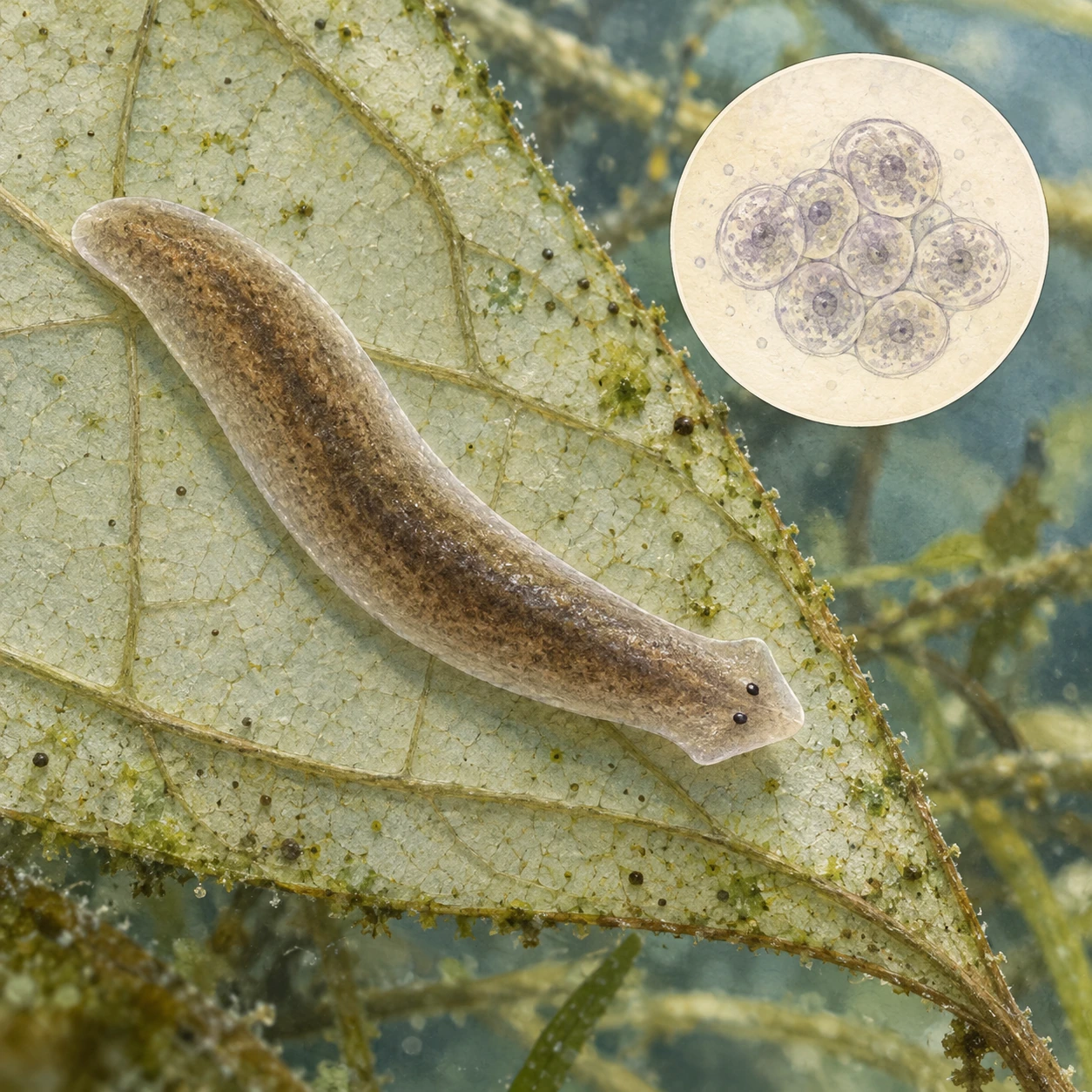
Intact planarian beside a conceptual enlarged cluster of round cells
A cell marker and a functional rescue answer different questions. The cell study is interpretive and is not microscopy or a mapped cell population.

A 2018 study combined single-cell gene-expression data with sorting and transplantation. A cell-surface marker, TSPAN-1, helped enrich for cells capable of rescuing the host after a single-cell transplant. This moved the identification partly from a retrospective question—what did a transplanted cell turn out to do?—toward prospectively selecting a useful population before testing its capacity.

A marker is not the capacity itself. Detecting a molecule can identify a promising class, while the transplantation outcome supplies a functional test. The distinction matters whenever a new cell type is announced from a gene-expression cluster alone.

Read source and setting as separate experimental parts

Sketch three boxes: donor cell, recipient body and descendants. Under the donor, ask how many cells were transferred and how they were selected. Under the recipient, ask what was altered and what tissues remained. Under the descendants, list which cell identities and functions were actually demonstrated.

Does one versatile cell contain a complete spatial plan?

The result does not establish that. It shows a cell's potential within the tested living context. The article's positional signals supply another part of the explanation: what descendants become and where they integrate cannot be reduced to merely having a source cell.

This is why the stem-cell and muscle-instruction discoveries fit together. One addresses the range of material the animal can generate. The other addresses the organization in which that material is used. A dramatic rescue experiment makes the capacity visible without making the surrounding body irrelevant.

A coordinate system can be distributed

Research on FGFRL–Wnt circuits examined regional expression in muscle and its role in organizing the head-to-tail axis. Genes with different spatial patterns can provide overlapping information. The system does not require one special cell that stores the location of every other cell.

The Whitehead account of the work explains how experimental manipulation helped reveal those functions. Changing a signal and observing a corresponding patterning change provides stronger causal evidence than finding that a gene is active near a structure. Location suggests a hypothesis; perturbation tests a role.

Even a causal experiment has boundaries. Suppressing a gene can affect several processes, and one observed outcome does not establish every interaction in the network. Researchers compare markers, tissues, timing, and controls to decide which interpretation best fits. The complicated part is not a distraction from the spectacle; it is how the spectacle becomes understandable.

Regeneration is also ordinary maintenance made visible

A 2022 synthesis connects positional information with stem-cell biology in regeneration and normal tissue turnover. Maintaining an uninjured adult also requires replacing cells without losing organization. Injury reveals an especially dramatic version of a continuing biological problem.

That connection avoids treating the worm as a bag of magical cells that activates only when damaged. The living body already coordinates cell production, differentiation, and position. Regeneration redirects those capacities in response to a changed condition.

It does not follow that any microscopic fragment of any flatworm can produce a complete animal, or that the same signaling intervention would give a human the same capacity. Species, fragment properties, and experimental conditions matter. The transferable scientific value lies in discovering general questions and testable mechanisms, rather than promising an immediate medical application.

The illustration shows intact animals so that the organism remains more than an injury demonstration. Its subtle internal pattern is interpretive. The paper's microscopy and gene-expression data provide the evidence for the cells and signals discussed here; no cutting experiment is needed at home.

A planarian's achievement is organized renewal. New cells are only part of it. Existing tissue supplies a changing context, muscle carries positional signals, and stem-cell descendants form appropriate structures. The small worm makes visible a large biological truth: to rebuild a body, growth has to become a pattern.

Follow the evidence: Reddien Lab: stem cells and positional information; Witchley and colleagues: muscle provides regeneration instructions; Scimone and colleagues: regional FGFRL–Wnt circuits; Reddien: positional information and stem cells in regeneration; Whitehead Institute: experimental body-patterning mechanisms.

More curious connections, art and independent projects at Incoprea.

Wednesday, October 14, 2026

How an Axolotl Rebuilds a Limb, and What Can Stop It

Closing a wound and replacing a limb are different achievements. An axolotl can do the second: rebuild a missing structure containing bone, muscle, skin and nerves in an organized arrangement. The remarkable feature is not simply that cells grow. It is that they contribute to a body part whose pieces must fit one another and the animal that remains.

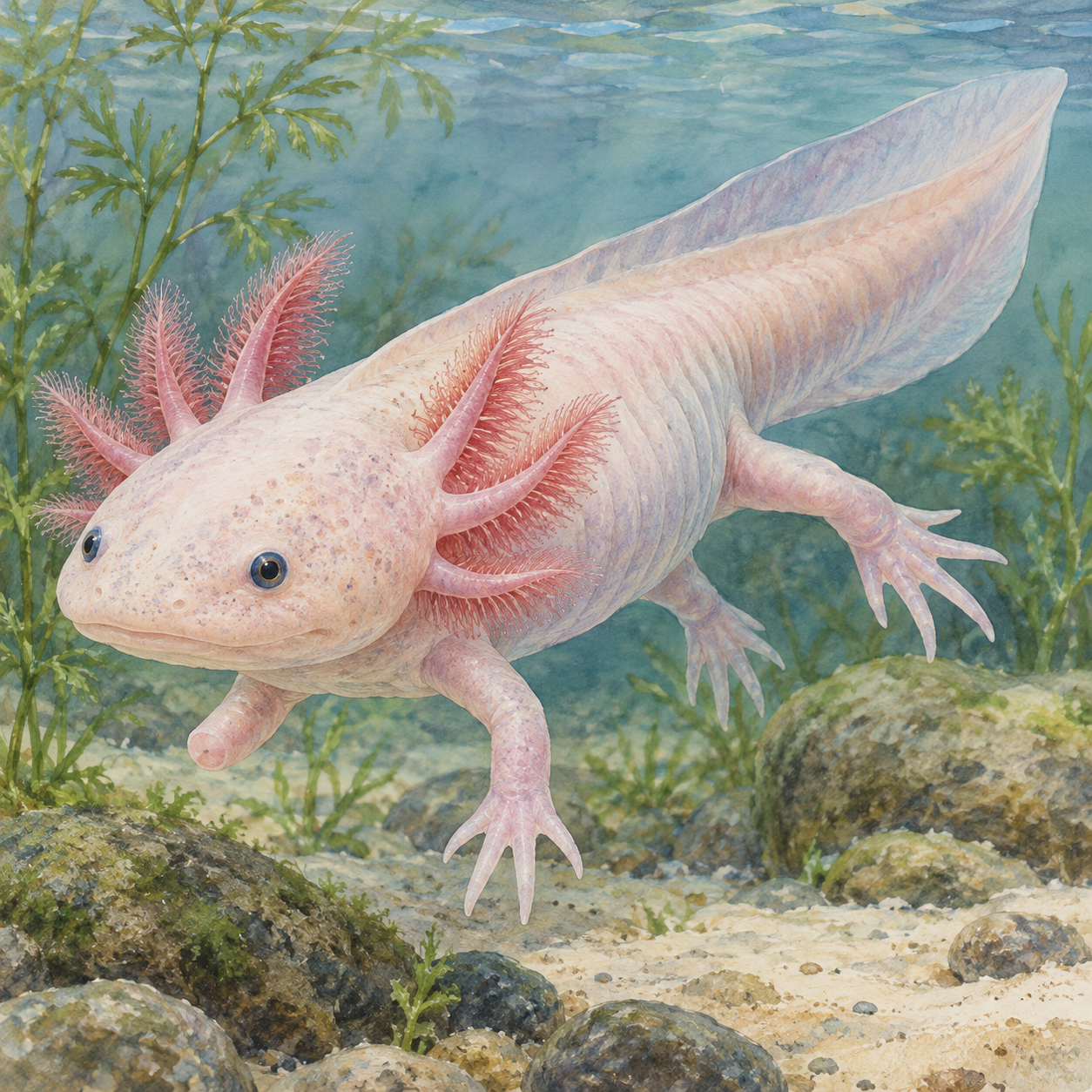
A pale axolotl with rose-colored external gills and a small forelimb bud swims in clear water.
An illustrative axolotl with a smooth limb bud suggesting regeneration, rather than a documented experimental specimen.

This aquatic salamander, Ambystoma mexicanum, offers researchers a way to study rebuilding as a sequence of events. A successful result shows what is possible. Interrupted or altered regeneration helps reveal what the sequence requires. Both are needed to move beyond the broad statement that an axolotl can grow something back.

Covering the wound is a beginning

After limb loss, a specialized wound epithelium forms over the site. Beneath it develops the blastema, a region containing proliferating progenitor cells. Signals associated with the wound covering, nerves and surrounding tissues help establish conditions for rebuilding. The site becomes a coordinated growth region rather than merely a sealed surface.

The blastema is often imagined as a bag of completely interchangeable cells. Martin Kragl and colleagues tested that idea by tracking labeled tissue lineages during regeneration. Their 2009 study showed that the blastema contains different populations with restricted contributions, retaining information about tissue origin. Regeneration did not require every participating cell to become capable of producing every tissue.

A useful analogy is rebuilding with several specialized teams rather than converting every worker into the same universal builder. The comparison should stop short of implying a foreman issuing instructions. The biological coordination arises through cells, signals and their interactions. Knowing where cells came from helps researchers understand how the different contributions are organized.

Making material is different from placing it

A regenerated limb needs more than the correct collection of cell types. Structures have positions: nearer the body or farther from it, on different sides, and in relation to joints and digits. Positional information is therefore part of the problem. Producing cartilage somewhere at the site is not equivalent to restoring an appropriately arranged skeleton.

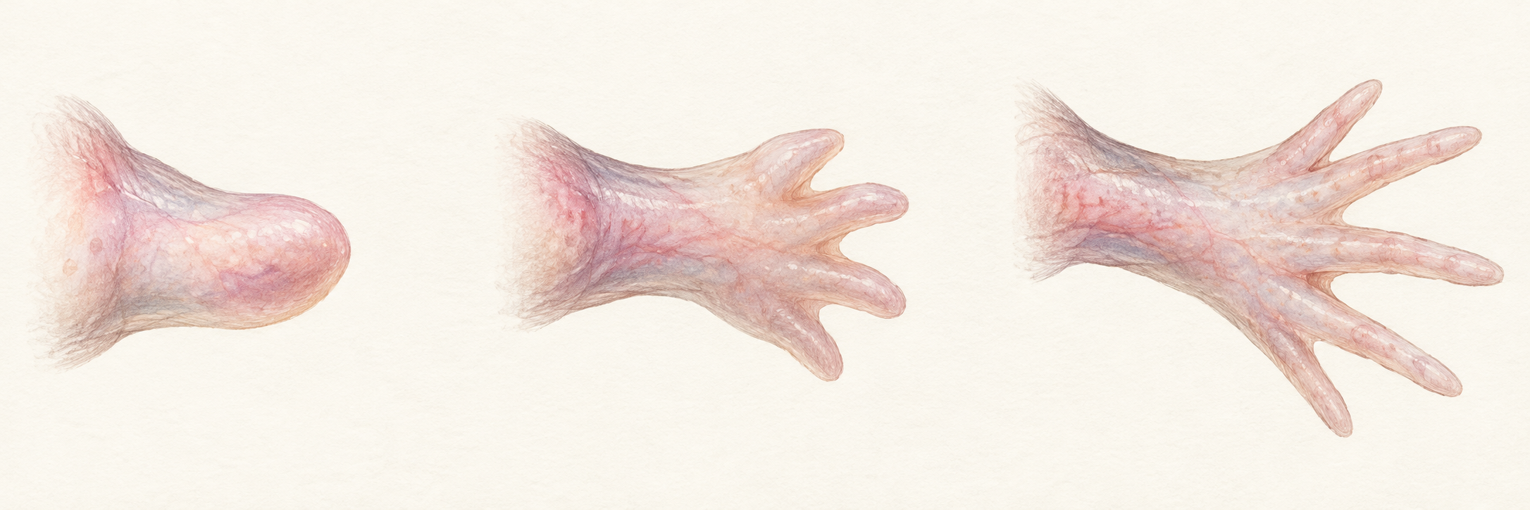
Use the supporting sequence as a guide to the broad transition from bud to limb, rather than a precise timetable. The illustration uses a hindlimb form; it is not a measured experiment. Actual studies can examine tissue sections, labeled cells and skeletal preparations to ask questions the outward silhouette cannot answer.

Three conceptual limb studies show a rounded bud, emerging digits, and a developed five-digit hindlimb form.
Conceptual studies of axolotl limb regrowth using a five-digit hindlimb form. The sequence is illustrative, with no exact timing or specimen scale.

Try evaluating a hypothetical photograph of a regrown limb. What would you need to know beyond its appearance? You might ask whether the internal tissues are arranged correctly, whether nerves connect appropriately, and whether the animal can use the structure. A recognizable shape is important evidence, but it is not the same measurement as normal function.

The immune response can help build the conditions

James Godwin and colleagues investigated macrophages, immune cells involved in responses to tissue damage. When the team depleted these cells during an early critical period, the wound could close but limb regeneration failed, with extensive fibrosis. The 2013 study separates surface repair from restoration of the missing structure in a particularly direct way.

That result also complicates the idea that regeneration succeeds simply by avoiding immune activity. The relevant cells and signals help create an environment in which rebuilding can proceed. Timing matters: removing a component during one part of the process can have a different effect from altering it later. “Inflammation” is too broad a word to identify every constructive or disruptive interaction.

Imagine comparing two repaired sites. Both are covered by skin, but only one develops the missing limb. If you recorded only whether the wound had closed, you would miss the central difference. A study's outcome measure determines which achievement it can detect. This is why regeneration research follows several stages rather than treating healing as a single yes-or-no event.

The state of the animal matters

Axolotls normally become reproductively mature while retaining features such as external gills. Researchers can study a different developmental state through experimentally induced metamorphosis. In a 2014 comparison, age-matched metamorphosed animals regenerated more slowly and produced more skeletal abnormalities than animals retaining the usual aquatic form.

Matching age helped separate developmental state from the simple fact of being older. The study also examined body size rather than assuming every large animal would behave like every small one. Such comparisons matter because several traits can change together. A dramatic before-and-after picture cannot identify which of those changes explains a difference in rebuilding.

The finding concerns a controlled research comparison, not an instruction to alter a pet's conditions. More broadly, it shows why the whole animal belongs in the explanation. A blastema does not operate independently of development, physiology and the tissues supplying it. The capacity to rebuild emerges from a biological setting, not from one isolated collection of cells.

Ask four questions of a regeneration claim

When you meet a new headline, ask whether the study showed wound closure, replacement tissue, correct pattern, or restored function. These achievements can overlap but are not synonyms. Then ask which organism and experimental conditions produced the result. The distinction makes it easier to appreciate a major finding without asking it to establish a different one.

NIH describes axolotls as valuable research organisms while noting that laboratory populations differ genetically from endangered wild populations. Learning how a captive research animal rebuilds a limb and protecting salamanders in their native Mexican waterways are connected by the species, but they are separate practical undertakings. A thriving laboratory colony does not substitute for a surviving wild habitat.

The axolotl's value is the opportunity to ask increasingly precise questions: which cells contribute, what information they retain, how the environment supports them, and why rebuilding sometimes loses speed or accuracy. Its successful limbs and its experimental limits illuminate the same process. Regeneration becomes a system to understand rather than a single miraculous ingredient to borrow.

Explore more creative curiosity at Incoprea.

The Sea Slugs That Keep Their Food's Solar Machinery

A green sea slug can look like a leaf that has begun to crawl. In some species, part of its meal really does continue catching light inside it. The slug eats algae, retains chloroplasts from their cells and keeps those structures photosynthetically active. Food has become more than fuel: some of its machinery is still working after ingestion.

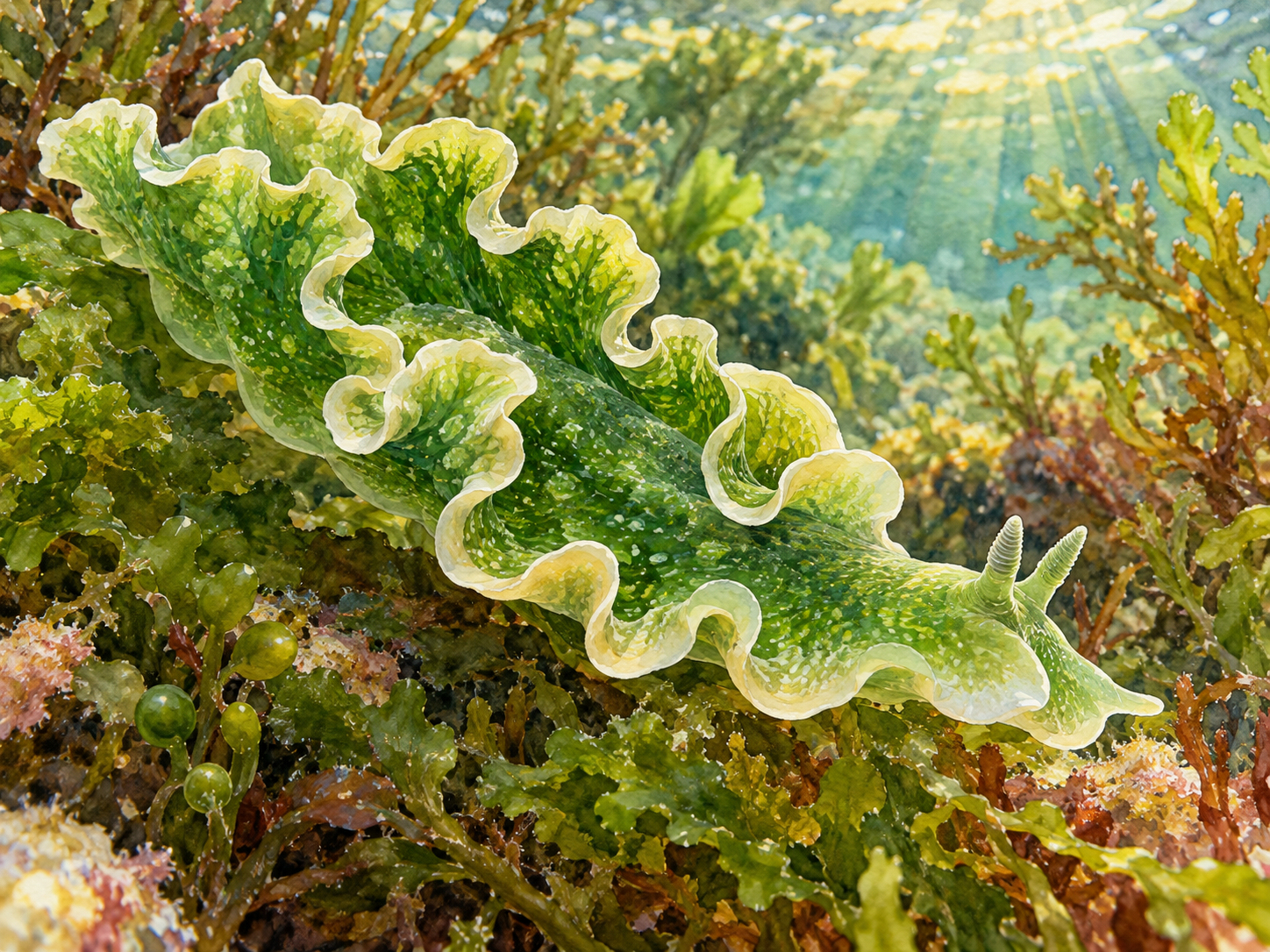
A green lettuce sea slug with ruffled pale-edged folds moves among marine algae.
A lettuce sea slug among algae, illustrating the animals associated with kleptoplasty.

This is kleptoplasty, the retention of stolen plastids. The animals involved are certain sacoglossan sea slugs, and the captured structures are chloroplasts, not entire miniature plants. That distinction creates the central mystery. A chloroplast ordinarily works within an algal cell that supplies support. How can it continue functioning after much of that original setting has been digested?

Follow what is kept and what is lost

Sacoglossans puncture algal cells and feed on their contents. In species capable of sustained kleptoplasty, some chloroplasts escape immediate digestion and are retained within cells associated with the slug's digestive system. The algal nucleus and other components of the meal do not simply remain as a complete alga living independently inside the animal.

A chloroplast has its own genetic material, but it normally also depends on proteins encoded in the algal nucleus. Removing it from that relationship is therefore more consequential than moving a self-contained solar panel. The panel analogy is helpful for light capture and misleading for maintenance. A living organelle needs an appropriate chemical environment and ways to withstand damage.

Sonia Cruz and Paulo Cartaxana's review emphasizes how much this differs among species. Some slugs retain chloroplasts briefly; others maintain activity for weeks or months. The relevant unit of comparison includes the animal, its algal donor and the conditions of the observation. A single impressive retention time cannot become a promise about every green slug.

The host builds a place for the machinery

A 2025 study led by Corey Allard investigated the lettuce sea slug, Elysia crispata. The team identified host-derived compartments surrounding captured chloroplasts, which they called kleptosomes. The name refers to the slug's surrounding organelle, not a replacement name for the chloroplast itself. There is a biological boundary around the retained machinery.

The researchers combined microscopy, protein analysis and recordings from organelle membranes. They identified ATP-sensitive ion channels associated with the compartment and evidence that disturbing this system reduced photosynthetic output under the tested conditions. The result makes the host an active participant in maintaining the captured chloroplasts, rather than merely a passive bag holding green material.

During prolonged starvation, the study found a shift toward degradation of stored chloroplasts. A compartment supporting retention can therefore also participate in eventual digestion. The useful machinery and the stored food are not necessarily competing explanations at all times. The host's state helps determine how the captured material is used.

Green is only the first rung of evidence

Imagine three observations. First, a slug looks green. Second, measurements show light-dependent photosynthetic activity. Third, newly fixed carbon appears elsewhere in the slug's tissues. Which observation would establish that useful products leave the retained chloroplasts? The third asks the most direct question about nutritional transfer. Color alone cannot supply that answer.

Think of those observations as a short evidence ladder. Presence of pigment suggests something worth investigating. Photosynthetic measurements establish a process. Tracing the products connects that process to other tissues. Each rung adds information, and none should be silently replaced by the one below it. A beautifully green photograph is not a carbon-tracing experiment.

Paulo Cartaxana and colleagues used isotope labeling and imaging to follow this transfer in Elysia timida. Light-dependent incorporation of labeled carbon was detected beyond the chloroplast-bearing digestive cells, including reproductive tissues. Controls without the relevant illumination helped distinguish photosynthetic incorporation from other possible routes. The study supplied a connection between retained organelles and the rest of the animal.

In another part of that work, animals under regular light produced more eggs than those under reduced light in the experimental conditions. The researchers considered remaining limitations, including possible direct effects of light on spawning. The result supports a reproductive benefit in the tested system without establishing that sunshine replaces all ordinary feeding in every species.

Useful light can also become a challenge

Photosynthesis does not mean unlimited exposure to light is always helpful. The light-processing machinery can be damaged when incoming energy exceeds what it can safely use. Photoprotection and repair are therefore part of keeping chloroplasts functional. A slug retaining the equipment must live with this balance, alongside the benefits of photosynthetic products.

A 2024 comparison of Elysia species found differences in photoprotective capacities, including contrasting responses in E. timida and E. crispata. That variation reinforces the importance of retaining species names in the story. A successful maintenance mechanism identified in one host does not establish identical performance or the same protective toolkit in another.

For a useful comparison, ask two questions of any “solar-powered animal” headline. What evidence shows photosynthetic activity, and what evidence shows a benefit to the animal? Then ask which host and donor were involved. Those details turn an appealing category into a biological relationship whose costs and advantages can actually be investigated.

Borrowing an organelle is a particular achievement

Chloroplasts have a much older evolutionary history involving photosynthetic bacterial ancestors and the cells that integrated them. A sea slug retaining chloroplasts within one lifetime is not replaying that entire history. The slug remains dependent on acquiring its algal meal and has not become a permanently independent, plant-like photosynthetic lineage.

Its achievement is distinctive enough: foreign machinery enters an animal cell, receives a regulated compartment and contributes to processes beyond the original meal. The leaflike appearance opens the story, but the most interesting action is at a boundary you cannot see unaided. Inside it, the animal regulates whether a useful part of its food is preserved or eventually digested.

Explore more creative curiosity at Incoprea.

Famous Last Meals is now in paperback — stories, history and 25 recipes

Famous Last Meals: Stories, History, and Recipes is now available in paperback on Amazon. The illustrated collection brings together 23 his...