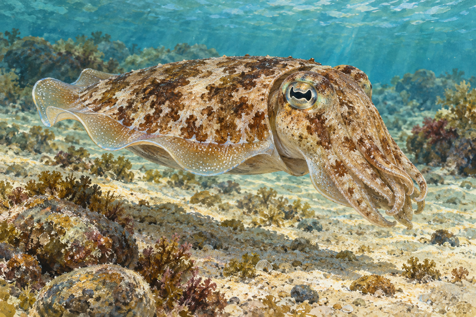
A cuttlefish can shift from a pale mottled appearance to strong dark markings while it hovers above the seabed. The transformation can look like an animation playing across its body. A central part of the effect is a set of skin organs called chromatophores.
Calling them pixels is a helpful analogy, provided we remember that each is living tissue. Muscles, nerves, elastic material and pigment work together to change how much colored surface is exposed.
A tiny sac that spreads into a disk
A cephalopod chromatophore contains an elastic pigment sac connected to muscles arranged radially around it. When those muscles contract, they pull the sac outward, spreading it into a broad colored disk. When the muscles relax, the sac retracts and its visible colored area becomes much smaller.
The pigment does not have to be manufactured afresh for every quick change. Mechanical expansion and contraction can expose or hide existing color. Coordinating many such organs creates patches, bands and complex body patterns.
These muscles are controlled through the nervous system. A cephalopod can therefore alter its appearance rapidly as it responds to its surroundings or other animals. The pattern is part of behavior, rather than simply a passive change in the chemistry of its skin.
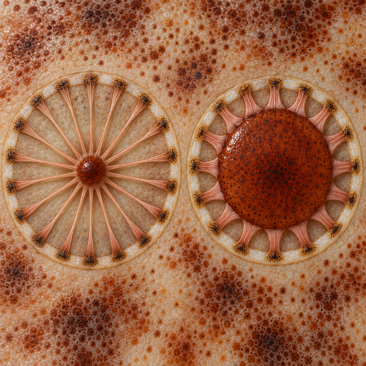
Expanding an existing color changes the visible pattern
A useful way to read the sac diagram is to choose one pigment organ and follow its edge. The colored area expands when the surrounding radial muscles shorten. This can seem backward if we imagine that muscle contraction must always make the visible feature smaller. Here, the muscles pull outward from different directions, spreading the central elastic structure.
The resulting disk changes how much pigment is exposed over a patch of skin. A small dark point can become a much broader dark region without a new supply of pigment arriving at that moment. Coordinated over many organs, changes in exposed area produce a body pattern that can transform much faster than a new coat of pigment could be built from scratch.
The “pixel” comparison captures spatial control, but a screen pixel is not a complete model. The animal's skin has layers and mechanical depth, and reflection changes with illumination and viewing angle. A similar color in two frames can arise through different combinations of pigment exposure and optical effects.
This is why the microscopy is especially useful. It lets us follow the same small structures across time instead of assigning one isolated image to one mechanism. Their changing outlines show a mechanical process, while the accompanying sparkle raises questions about light reflection.
More than one way to make color
Chromatophores are important, but they are not the whole optical system. Cephalopod skin also includes reflective structures. Iridophores produce iridescent effects through microscopic organization, while leucophores can contribute pale or white reflection. The combination gives the animal more possibilities than a layer of pigment alone.
The division is not always as tidy as an introductory diagram suggests. A 2019 study of squid chromatophore organs reported structural coloration associated with the organs themselves, including reflectin proteins in cells around pigment sacs. The researchers found that pigmentary and reflective effects could be intertwined.
This matters when interpreting a shimmering blue or changing sheen. It may depend on how light is reflected, including the viewing angle, rather than on a blue pigment expanding in exactly the same way as a brown one. Species and tissues differ, so a result from one squid should not automatically be treated as the precise mechanism of every octopus display.
Watch the outline first, then the sheen
On the first viewing, choose a dark or yellowish patch and watch its boundary. Does its area change, or does its brightness change while the outline stays broadly similar? Do neighboring patches move together? You are looking for changes that a simple statement such as “the squid turned blue” would hide.
On the second viewing, follow the reflected glints. Notice how their appearance relates to the lighting and the changing tissue. The film is not a claim that every shimmer is produced by a separate blue pigment sac. The 2019 study examined structural coloration associated with chromatophore organs, complicating a tidy division between pigment above and reflectors below.
Finally, compare the microscope view with the cuttlefish scene. The larger animal's outline, posture and skin texture contribute to what a viewer or predator sees. The close view explains a component; it does not reproduce the whole behavior. A cephalopod's remarkable display emerges when these scales are put together, with each observation kept attached to the species and tissue actually studied.
Camouflage is a whole-body performance
Color and pattern can help an animal blend with its setting, interrupt its outline or communicate. Some cephalopods also change posture and skin texture. Cuttlefish papillae, for example, are muscular structures that can make the surface appear more three-dimensional. They are distinct from the pigment organs.
For a second microscopy view, MBL's research page includes microscopy videos of squid skin.
Sources and further reading: MBL: Squid skin coloration strategies · Dynamic pigmentary and structural coloration within chromatophore organs · MBL: How cuttlefish change skin texture