Showing posts with label Marine Life. Show all posts

Sunday, October 25, 2026

The Microscopic Thread That Turns Inside Out to Sting

A sea anemone's tentacle can look soft and uneventful. At its surface, however, specialized cells contain compact structures ready for an extraordinary transformation. A thread that was packed inside a capsule can discharge and turn inside out.

“Tiny harpoon” captures the suddenness, but misses much of the mechanism. The apparatus changes shape as it operates. Its unfolding architecture is part of how it works.

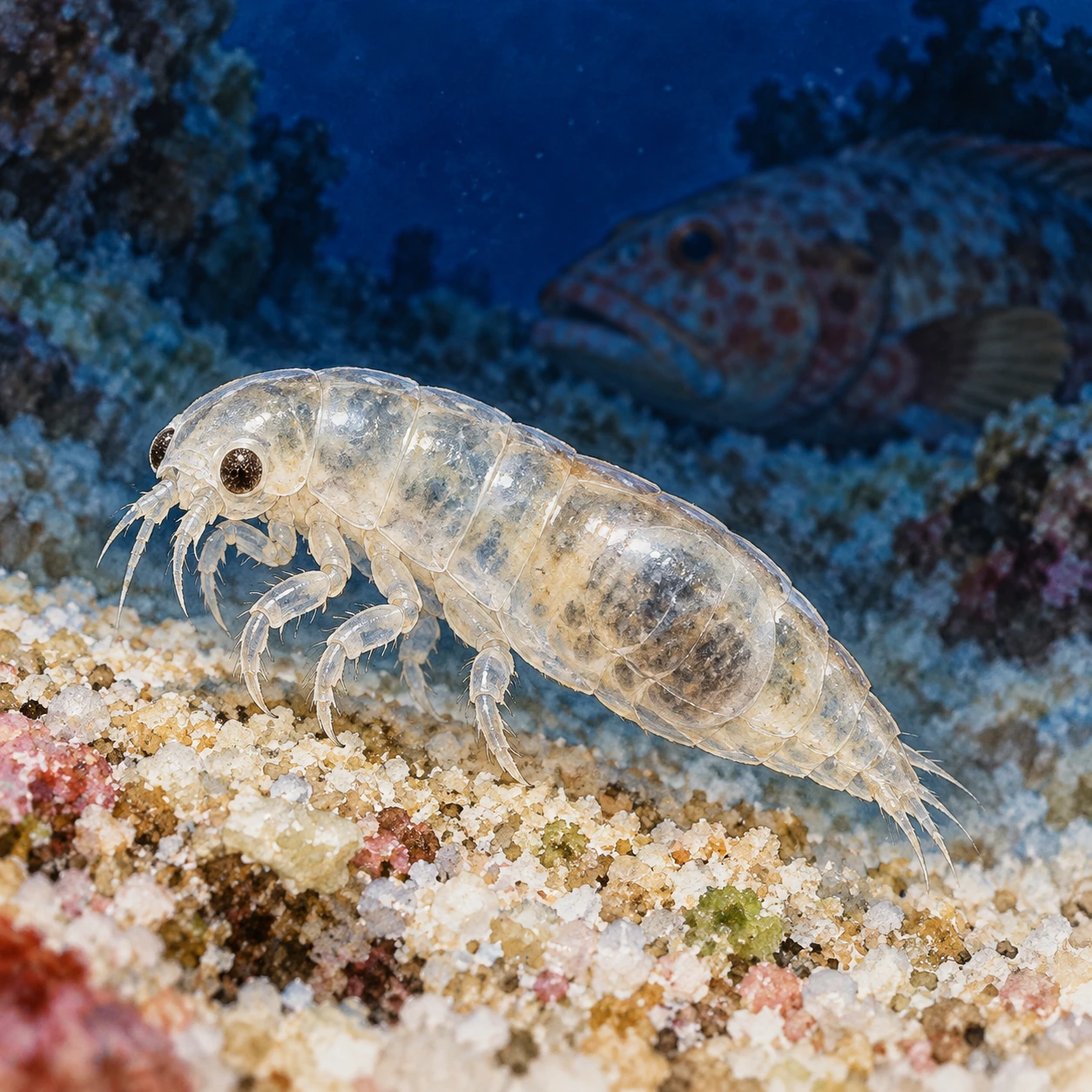
Small translucent hydroid polyp with tentacles extended from a seagrass blade.
Cnidarian tentacles carry specialized cells whose microscopic capsules can discharge an everting thread.

A cell containing a single-use capsule

Cnidarians include anemones, corals, jellyfish, and hydroids. Their specialized cnidocytes contain different kinds of capsules. The stinging capsules called nematocysts are one part of this diverse equipment.

The capsule and the cell are different levels of organization. A nematocyst is an organelle within a cell, rather than a free-living creature. Its stored thread and capsule must first be built by biological processes before discharge becomes possible.

The Stowers Institute's account of its researchers' work describes a system combining osmotic pressure with elastic energy stored in several substructures. Pressure supplies a powerful start, while the organization of the packed apparatus contributes to its subsequent transformation.

That arrangement separates preparation from action. A cell invests in making and loading the structure. When a suitable trigger arrives, it can release stored energy much faster than it could construct a new apparatus on demand.

Three phases, rather than one flying object

A 2022 study of the anemone Nematostella vectensis combined imaging methods to reconstruct the organization of a particular nematocyst type. Its researchers identified three main phases: shaft discharge, shaft eversion, and tubule eversion.

First, a tightly packed shaft is expelled. Next, its filaments uncoil and turn inside out, producing an elongated passage. The thinner tubule then everts through that passage. In this nematocyst, the shaft is both an initial penetrating structure and a protective route for the more delicate tubule.

The authors used three-dimensional electron microscopy alongside recordings of discharge. Their reconstruction links what was packed inside to what appeared outside. It is a mechanism for the studied structure, rather than an assertion that every cnidarian capsule has identical internal parts.

That specificity is valuable. Different nematocysts can perform different tasks, and comparing their architectures helps explain why a single familiar metaphor cannot stand in for the entire group.

Which part was extraordinarily fast?

The speed deserves careful reading. In 2006 research on Hydra stenoteles, specialized high-speed imaging resolved an initial discharge phase on a timescale as short as 700 nanoseconds. That is less than a millionth of a second.

The number describes an exceptionally rapid stage in the studied system. It should not be attached to the complete sequence in every anemone or jellyfish. The 2022 work explicitly distinguishes the faster Hydra mechanism from the slower shaft eversion it observed in Nematostella.

Whenever a biological speed sounds unbelievable, ask what began the stopwatch and what stopped it. Initial acceleration, full extension, and the animal's entire response are different measurements. The distinction preserves the achievement rather than diminishing it.

Why it does not fire at every disturbance

A one-use capsule needs more than an efficient launch. It also needs appropriate control.

A 2020 study of Nematostella investigated a specialized voltage-gated calcium channel in its stinging cells. The channel's properties suppress responses to irrelevant mechanical disturbances. Prey-related chemical signals can relieve that inhibition, allowing a mechanical stimulus to help trigger discharge.

This is sensory filtering at the cellular level. The animal lives in moving water; movement alone would be an expensive reason to expend every capsule. Integrating cues helps connect the available apparatus to a biologically relevant encounter.

Even that control is not one universal rule. Later research comparing Nematostella with Exaiptasia diaphana found differences in channel behavior and stinging responses. The comparison associated more tightly filtered predatory responses with one system and more readily activated defensive responses with the other.

The full story therefore has two intertwined achievements. A cell prepares a structure capable of rapid mechanical change, and sensory machinery helps determine when that structure is used. The quiet tentacle holds both a loaded geometry and a decision about contact with the world.

Read the research

Keep exploring

Discover more art, ideas, and curious things at Incoprea.

Thursday, October 22, 2026

Why Sea Otters Can Protect a Kelp Forest Without Rebuilding the Next One

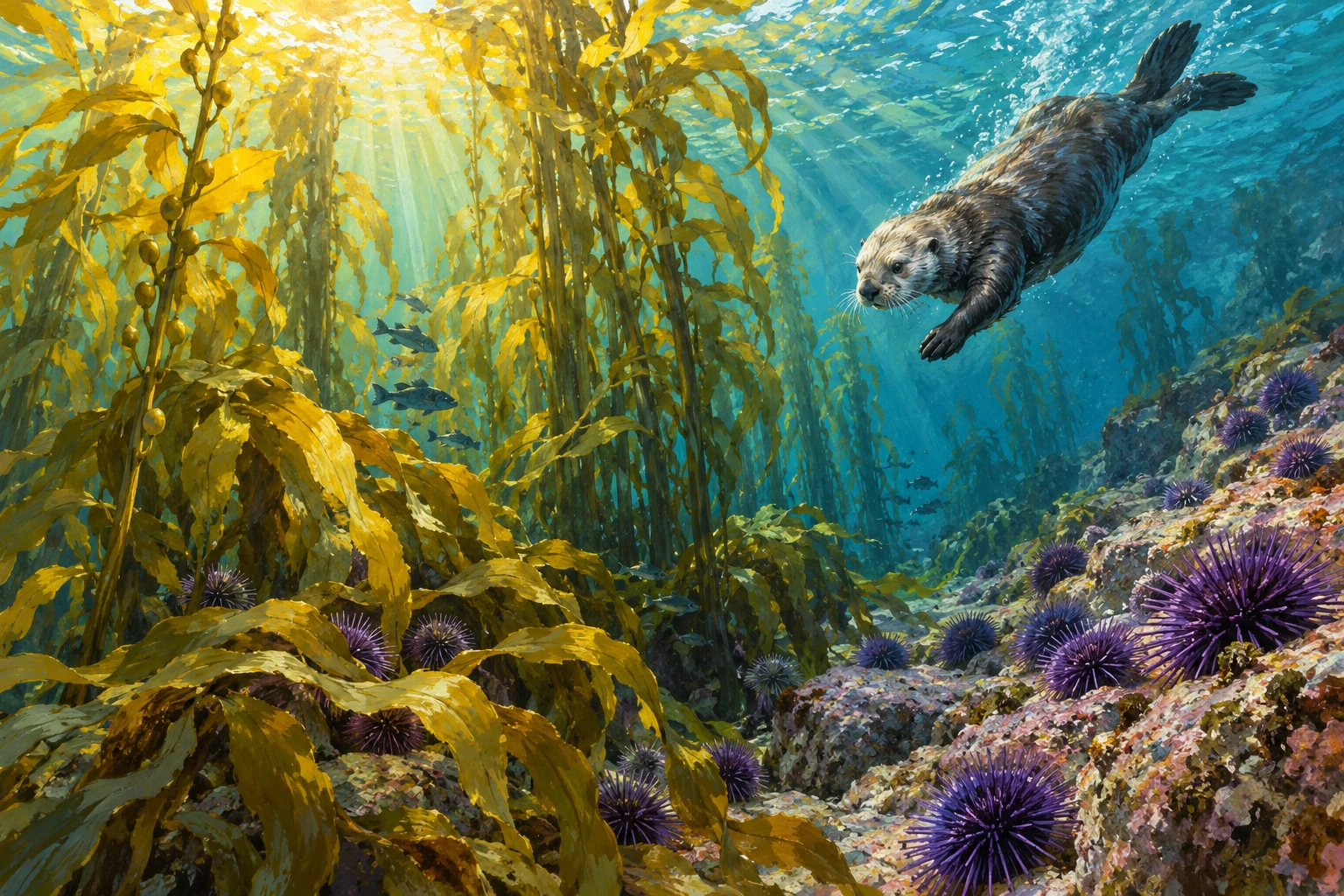
A sea otter dives toward the edge of a kelp forest beside rocky habitat dotted with purple urchins.
Conceptual ecological painting combining contrasting habitat patches; not a survey image or map.

The familiar kelp-forest story fits neatly into three links: sea otters eat sea urchins; sea urchins eat kelp; otters therefore help kelp forests persist. It is a real and influential example of a trophic cascade. But imagine seeing healthy kelp beside an urchin-covered barren while otters remain in the same bay. The next question is not whether the textbook was entirely wrong. It is what those three links left out.

A predator does not consume an abstract unit labeled “urchin.” It encounters individual prey with different nutritional value in different places. An urchin does not always graze in the same way. It may remain sheltered while drifting food reaches it, or emerge when that supply declines. Once behavior and prey condition enter the picture, the food chain becomes a landscape of choices and feedbacks.

A useful first diagram, followed by a harder question

NOAA's aquatic-food-web introduction uses otters to explain how a predator can have indirect effects on organisms it does not eat. More grazing pressure can reduce the kelp that supplies structure and habitat. Reducing grazers can therefore have consequences well beyond the predator's immediate meal.

The diagram is a starting model. It directs attention to a mechanism and predicts a possible response. It does not say every kelp forest has the same species, temperature, disturbance history, or resource supply. It also does not specify whether the predator will spend equal effort in every patch after the ecosystem changes.

That distinction became especially useful in Monterey Bay. A 2021 PNAS study examined the mosaic of kelp patches and urchin barrens. Researchers combined underwater surveys, otter foraging observations, and long-term ecological information. Rather than treating otter presence as a simple yes-or-no variable, they asked how the animals responded to different states of the habitat.

Many urchins can make a poor buffet

The UC Santa Cruz account of the research describes a crucial result: urchins in remaining kelp forests could be more rewarding prey, while urchins in barrens were often nutritionally poor. Otters concentrated feeding where the return was worthwhile. This could help defend existing forest patches without automatically clearing neighboring barrens.

It is easy to see the mistaken assumption in hindsight. A patch carpeted with prey looks like an abundant food resource to a distant observer. But abundance is not equivalent to edible energy. An animal foraging for a living must encounter, capture, process, and digest food. The visible number of prey is only part of the return on that effort.

Now the persistence of the mosaic makes more sense. In a forest patch, otter predation can limit exposed, rewarding urchins. In a barren, hungry urchins may remain abundant while being unattractive to otters. The same predator can contribute to the survival of one state without readily reversing another. Maintaining a forest and restoring a forest are related but distinct ecological outcomes.

The average otter can be an animal that does not exist

A coast's otters may consume many prey types without every individual eating the same mixture. That distinction adds another layer to the patchwork of kelp and barrens. Knowing the population is present still leaves the question of which particular animals are doing the foraging.

In 2008 research comparing food-rich and food-poor conditions, greater population-level dietary diversity under food limitation was largely associated with differences among individuals. Otters specializing on particular prey also differed in related foraging behavior and handling efficiency. The population's broad menu did not simply mean every animal had expanded its own menu equally.

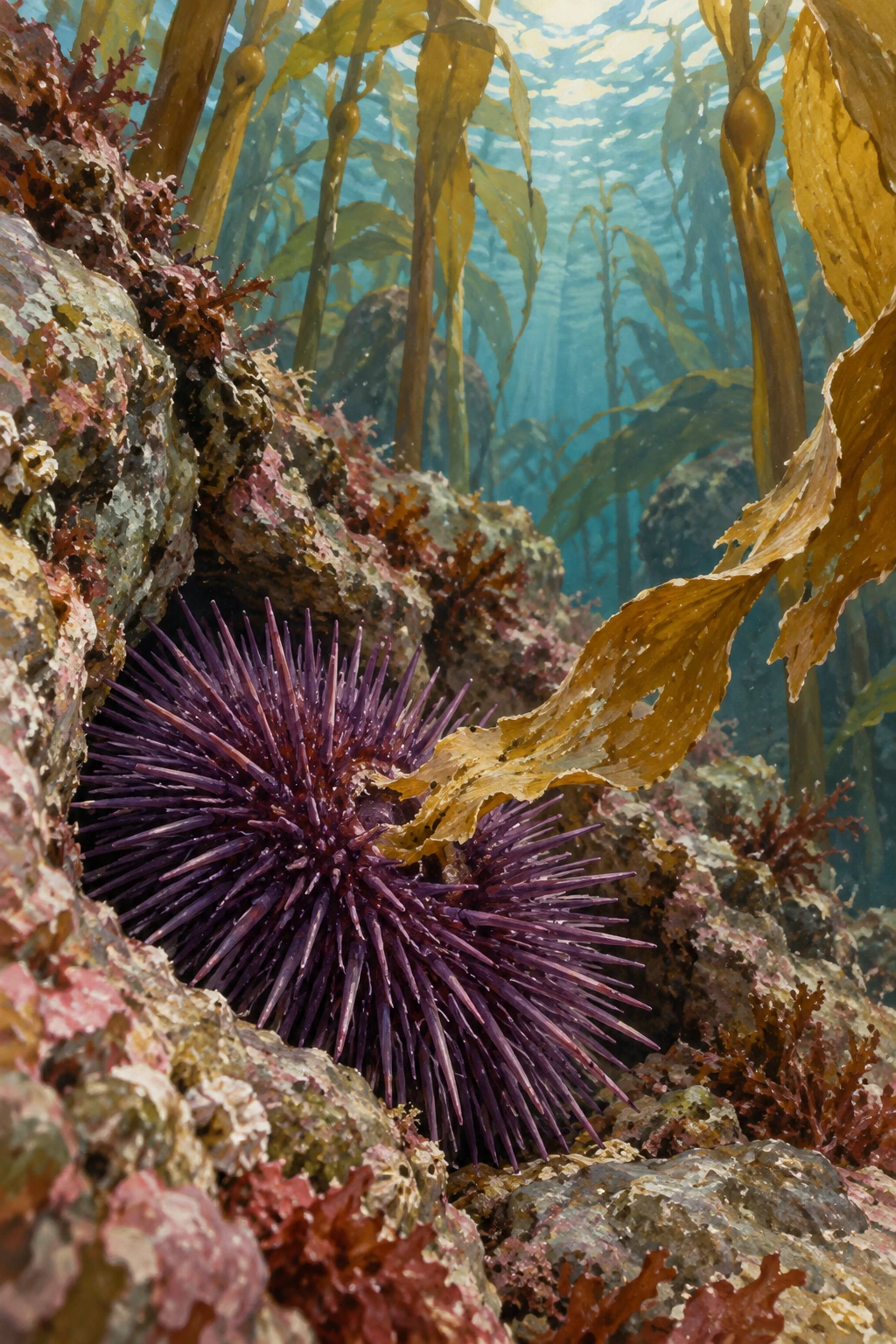
Purple sea urchin sheltered in a crevice near a drifting kelp fragment
An urchin occupies a particular feeding setting. The predator encountering it also has an individual foraging history.

The USGS record of the study describes the comparison made possible by translocation and long-term records of recognizable individuals. Following an otter repeatedly matters because an isolated meal cannot distinguish a persistent specialization from an opportunistic choice.

Try two fictional observation books. Each records 100 meals from five otters. In Book A, every otter supplies 20 meals of one prey type: one animal takes only crabs, another only urchins, and the remaining three each specialize on a different prey. In Book B, each animal takes four meals of each of the five prey types.

Pool either book and every prey type represents 20 percent of the 100 meals. Yet the individual stories are entirely different. The pooled chart cannot tell us which book produced it. It follows that a population diet graph alone cannot establish how any one otter will respond when a particular prey declines.

One extra column changes the question

Add individual identity beside every fictional meal. Group records by animal before calculating the pooled average. Then add date and observation effort. You can now ask whether preferences persist and whether apparent specialists were simply watched in different places or seasons.

This exercise does not explain the kelp mosaic by itself, and the invented meals are not survey data. It shows why behavioral diversity deserves a place beside prey abundance and prey quality. A rich ecological account can need several scales at once: the contents of one urchin, the history of one forager and the condition of an entire reef.

When the next restoration report describes “otter predation,” look for how it was measured. The same count of predators can conceal different diets, different skills and different distributions of effort across the coast.

Context is something to measure

A 2025 study of two long-term datasets compared kelp-community changes during sea-otter recovery at Vancouver Island and San Nicolas Island. Its focus was the dynamic, context-dependent strength of the cascade. The comparison is valuable because a mechanism can operate differently across places and through time. “Keystone species” does not mean a species produces one identical outcome wherever it arrives.

Long-term monitoring is what makes those comparisons possible. The USGS San Nicolas Island report describes population, foraging, and habitat surveys. These answer complementary questions. Counting otters tracks a predator population. Watching dives and identifying prey investigates feeding. Subtidal surveys record the community in which that feeding occurs. None can substitute entirely for the others.

For a reader following restoration news, a practical set of questions follows. Is a report describing a remaining forest, a recovering forest, or a barren that has not recovered? Did it measure prey quality and behavior, or only abundance? Is the evidence a short before-and-after comparison or a sustained record? Those questions do not dismiss a hopeful result; they help identify what the result can support.

The painting accompanying this article brings a kelp patch and an urchin-rich rocky patch into one conceptual view. It is an invitation to see a mosaic, not a map of a particular survey site. Real surveys attach places, dates, depths, and methods to such contrasts.

The sea otter remains a powerful ecological actor. The richer story explains its power more accurately. An otter can alter the future of a forest through repeated decisions about dinner, while the forest's condition changes what counts as a worthwhile dinner. The relationship is a loop, unfolding through individual animals and particular patches of coast. That is why the three-link diagram is memorable—and why the living coast rewards a second look.

Follow the evidence: NOAA: direct and indirect effects in aquatic food webs; Smith and colleagues: behavioral responses across kelp and barrens; UC Santa Cruz: otters maintain remaining kelp patches; 2025 study: context-dependent keystone effects; USGS: San Nicolas Island monitoring and foraging surveys.

More curious connections, art and independent projects at Incoprea.

Whale Falls: The Communities That Inherit a Body

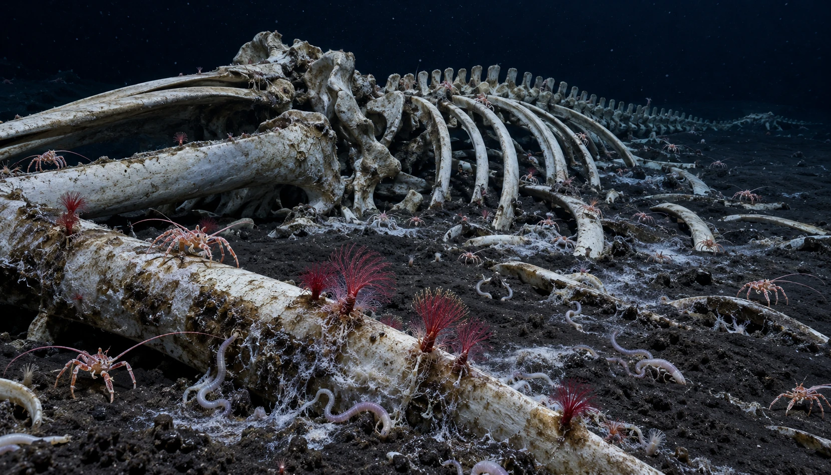
A whale skeleton on deep-sea mud with small crustaceans, worms and red Osedax plumes on a foreground bone.
Conceptual late-stage whale-fall community; organisms and visibility are arranged for explanation, not a recorded dive.

A whale skeleton on the deep seafloor can look like the final frame of a story. For the animals around it, the skeleton belongs to a changing opportunity. Flesh, bone, oil, microbes, and chemically altered sediment provide different resources on different schedules. A whale fall is therefore both a large meal and a small landscape with a history.

The most useful question is not simply “what eats a dead whale?” It is “what becomes available as one group feeds, and who can use what remains?” An animal that can tear soft tissue from a fresh carcass faces a different task from a worm living in a bone months or years later. Decomposition does not erase biological structure all at once. It changes which structures matter.

A succession of resources

The Smithsonian's illustrated whale-fall film introduces the familiar stages. Mobile scavengers first consume soft tissue. Smaller animals exploit organically enriched remains and surrounding sediment. Later, microbial processes associated with the remaining organic material can support communities using reduced sulfur compounds. The stages describe changing opportunities, not a universal clock that every carcass follows precisely.

That last point matters when an image circulates without a date. A bare skeleton does not necessarily mean the site has become biologically unimportant. Conversely, a dramatic carpet of animals need not be a permanent community. Depth, whale size, the condition of the carcass, local organisms, and the rate of decomposition can influence the sequence.

The research synthesis by Smith and Baco established whale falls as ecological systems worth studying across those stages. It also framed questions about connections with other deep-sea habitats. A carcass is isolated to a human observer looking through a vehicle camera, but colonists can connect it to a wider ocean through dispersal and shared habitat requirements.

The bones are still food

One of the most surprising residents is Osedax, a genus of bone-eating worms. The visible plume is only part of the animal. Root-like tissues extend into bone, and microbial associations help the worm use the resource. MBARI's research program follows these animals and the communities associated with them. Their feeding is a reminder that “picked clean” is a judgment made from the perspective of an animal interested in flesh.

Bone is not merely a mineral scaffold with no nutritional possibilities left. Materials locked within it remain accessible to specialized biology. Access changes the habitat in turn: organisms that penetrate and break down bone can affect how long it remains available to themselves and others.

Discovering the worms also changed estimates of diversity. MBARI's account of subsequent research describes a growing collection of distinct Osedax species after the first discovery. What initially looks like one kind of red fuzz can hide multiple evolutionary lineages. A distant camera view and a genetic identification are different kinds of evidence.

An island does not have to contain only island specialists

Because whale falls are unusual, it is natural to imagine every resident as a whale-fall specialist. MBARI research using five experimentally placed carcasses found a more mixed picture: many animals also occurred in other deep-sea environments, while some abundant residents were particularly associated with bones. A habitat can be distinctive because of the density and combination of its inhabitants, even when many species are not exclusive to it.

Experimental placement helps answer questions that a chance discovery cannot resolve as easily. Researchers know something about the starting material and can revisit it. Yet a placed carcass is still one set of circumstances, not a substitute for surveying the range of naturally occurring falls. Combining controlled opportunities with observations across different places produces a stronger account.

This changes how to read the common “island of life” metaphor. It captures the concentrated resource, but can conceal connections. The whale's body accumulated material during its life in the wider ocean. After death, dispersing organisms find it, microbes transform it, and some residents may use other habitats between such opportunities. The island belongs to a network, even when the camera shows only darkness beyond its edge.

The generated panorama here compresses a late-stage community into an explanatory scene. It is not a record of a single dive, and the small organisms are made legible for the reader. For genuine close-up footage and the full sequence, the linked research organizations provide a better next step.

The whale's end is real. So is the beginning of a series of other lives around its remains. What makes a whale fall compelling is not a comforting promise that death disappears, but the specific work of transformation: one body becomes different resources, and each resource opens a different biological door.

Follow the evidence: Smithsonian: whale-fall succession and film; MBARI: experimental whale falls and community overlap; MBARI: Osedax studies and decomposition; MBARI: diversity of bone-eating worms; Smith and Baco: ecology of whale falls at the deep-sea floor.

More curious connections, art and independent projects at Incoprea.

A Parrotfish’s Mucus Cocoon Is a Barrier With a Specific Job

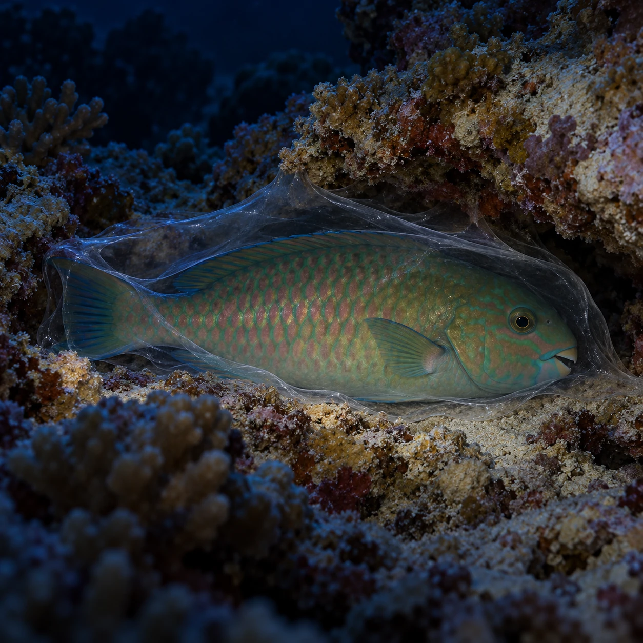
A parrotfish rests in a reef crevice within a thin nearly transparent mucus envelope.
Conceptual resting scene; the envelope is interpretive, not an experimental specimen or a measured barrier.

At night, a reef fish can settle inside a covering almost too clear to see. The cocoon is made of mucus, secreted around the resting animal. It looks vulnerable compared with a shell or a burrow, yet for some parrotfishes it can provide a useful barrier against small blood-feeding parasites.

The comparison often used is a mosquito net. Like any analogy, it identifies a function without establishing an identical mechanism or construction. The fish does not weave cloth. It produces a physiological covering whose value can be tested by asking what happens when the covering is absent.

The experiment behind the familiar image

A study published online in 2010 and in a 2011 issue of Biology Letters investigated mucous cocoons in Chlorurus sordidus. Researchers compared fish that retained their cocoons with fish from which the cocoon had been removed, and examined attack by gnathiid isopods. These small crustaceans feed on fish blood during part of their life cycle.

In the reported experimental comparison, 94.4 percent of fish without their cocoon were attacked, compared with 10 percent of fish that retained it. This was a defined test with particular fish, parasites, timing, and conditions. It supports protection against those attacks; it does not establish that a cocoon makes a fish invulnerable to everything in the reef.

The Smithsonian's photograph and caption provide a visual reference for the covering. The genuine cocoon is a delicate envelope, not a rigid transparent sphere. Seeing its thinness makes the experimental question more interesting: the barrier does not need to look like armor to alter access for a tiny parasite.

Why removal is more informative than a photograph

A fish in a cocoon demonstrates an association between a behavior and a material structure. To establish a protective function, researchers need a relevant comparison. Removing the structure while preserving other conditions as well as possible changes the proposed cause and lets the outcome be measured.

Even then, the design deserves inspection. Handling could change the fish's state. Some fish might abandon or damage a cocoon or make another. The published methods specify how such cases were treated. Those details determine which animals the final numbers describe.

This is a useful reading habit for any natural-history experiment. Ask what changed intentionally, what else might have changed with it, and which observations were excluded. A clear result becomes more convincing when its construction is visible, not when the procedure is omitted for a cleaner story.

A parasite count is the balance of several movements

The number of parasites on a fish at one instant is not the same as the number that tried to feed during the night. New arrivals, successful attachment, departure after feeding and removal can all change the count. A protective covering can alter one part of that traffic.

Enlarged interpretive gnathiid larva on reef sand with a distant fish
A small parasite’s arrival, feeding and departure belong to different stages. The illustration is not microscopy or an identification key.

A 1999 study of the wrasse Hemigymnus melapterus compared gnathiid abundance and feeding state through the day. More parasites were present at dawn than at sunset, but the evidence also indicated new infestations during daylight. This concerned a different host from the parrotfish cocoon experiment. It shows why time of sampling belongs in the biological explanation.

2002 work on cleaning interactions then combined observations with a model of parasite arrival, departure and removal. The clients' cleaning behavior did not simply track every daily change in parasite abundance. A count could vary while the frequency and duration of inspections remained comparatively steady.

Try a balance sheet with a missing history

Begin with ten fictional parasites on a fish. In Scenario A, five arrive and five leave during an interval. In Scenario B, none arrives and none leaves. Both finish at ten. The same final count describes very different exposure histories.

What if a barrier prevents four arrivals?

Under the invented A schedule, one arrives and five leave, so six remain. Under B, there were no arrivals to prevent and ten remain. The difference does not make the barrier intrinsically better in one fish; its observed effect depends on the traffic occurring during the interval.

These fictional cases omit feeding success, parasite identity and possible host responses. They isolate one measurement issue: a standing burden is an outcome of rates through time. The arithmetic is not a reconstruction of the cocoon study.

Return to the removal experiment with that distinction in mind. Ask when exposure began, when attacks were counted and how the intact and removed treatments shared the interval. The result becomes a precise test of protection under specified circumstances, rather than a timeless claim that a fish is either parasitized or protected.

A delicate covering can matter because it changes access during a vulnerable period. The small animals outside it are part of a moving system, not a fixed set of dots waiting on the skin for the observer's next photograph.

Making the covering has a cost

The same cocoon study estimated energetic investment using the material's energy content and a daily assimilation estimate from another parrotfish species as a proxy. Its often repeated figure of roughly 2.5 percent is therefore an estimate with a cross-species assumption, not a complete direct measurement of every cost in the study fish.

That qualification is biologically interesting. A protective behavior can be worthwhile when its benefits exceed its costs, but identifying those costs accurately is difficult. Material energy, secretion, time, and alternative activities are not automatically captured by one number. The calculation offers a starting comparison, not the final accounting.

Some fish also produced a second cocoon after removal. That shows a capacity to respond within the night under the study conditions. It does not mean repeated removal would be harmless, or that a diver should touch a resting fish to see the response. Observation should leave the covering intact.

A barrier against one parasite is not a universal parasite shield

A 2020 study of trematode infections in Okinawan parrotfishes considered a different group of parasites. Cocoon use did not straightforwardly explain prevalence across the species examined. That does not contradict the gnathiid experiment: different parasites reach hosts in different ways, and comparisons across species introduce additional differences.

The two studies illustrate why a general statement such as “mucus prevents parasites” is too broad. A useful account identifies the parasite, how it encounters the host, which host species is involved, and what was measured. A mechanism can work well within one interaction and be irrelevant in another.

The night reef also differs from the daytime reef familiar to snorkelers. Rest changes what a fish is doing, and a covering can help alter exposure during that vulnerable interval. The seemingly quiet scene contains ongoing interactions among a resting vertebrate, secreted material, and small organisms searching for a meal.

The generated painting here suggests the envelope without pretending to reproduce an experimental specimen. The linked Smithsonian image is the better visual evidence for the real covering. Taken together with the experiment, it changes what a bedtime scene can mean: a fish has made a temporary boundary between its skin and a particular kind of hungry neighbor.

Follow the evidence: Grutter and colleagues: fish mucous cocoons and parasite attack; Smithsonian Ocean: parrotfish in a mucus cocoon; Parrotfish trematode prevalence in Okinawa: a different parasite question.

More curious connections, art and independent projects at Incoprea.

Wednesday, October 14, 2026

The Small Lobster Nerve Circuit That Helps Explain Big Brains

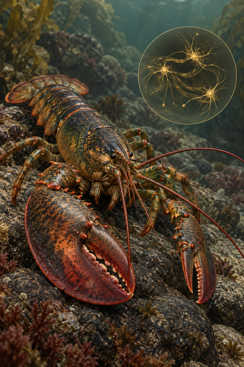
An illustrated lobster appears beside a small abstract inset of connected neurons.
Illustration of a lobster with a conceptual neural inset. The inset does not represent the actual ganglion location, cell count or connection map.

A lobster's digestion involves coordinated movements inside its body: food is chewed, filtered and moved along. Controlling those movements requires rhythmic signals. A small part of the crustacean nervous system has become one of neuroscience's most informative models precisely because researchers can study its components in detail.

The stomatogastric ganglion contains only about 30 neurons in the lobsters and crabs commonly used for this research. That is a particular ganglion, not the animal's entire nervous system. Its manageable size gives scientists a way to connect individual cells with the patterns they collectively produce.

A circuit that generates rhythms

Neural networks known as central pattern generators can produce repeated motor patterns. In the stomatogastric system, interacting circuits organize different aspects of digestive movement. The gastric mill circuit helps coordinate chewing, while the pyloric circuit organizes faster filtering and related movements.

Studied patternDigestive role
Gastric mill rhythmCoordination of chewing movements
Pyloric rhythmCoordination of filtering and pumping-related movements

Researchers can identify neurons, record electrical activity and investigate their connections. Under suitable laboratory conditions, an isolated ganglion can continue producing rhythmic activity. This makes it possible to study how a pattern arises without having to infer everything from the animal's visible behavior.

A rhythm is still influenced by the cell's properties, its connections and incoming modulatory signals. The network is not simply a tiny rigid clock that ticks identically in every circumstance.

Rhythm includes a sequence, not just a speed

Tap three fingers in a repeating order: first, second, third, then begin again. Now repeat faster while keeping the same relative spacing between the taps. The frequency has changed, but the pattern within each cycle remains similar. This limited analogy helps distinguish two properties of a rhythmic motor system.

In the pyloric rhythm, researchers can examine when different neurons become active relative to the cycle. A useful pattern is not defined only by how many bursts occur each second. The order and relative timing help organize the motor output. Two recordings with different overall speeds can therefore preserve an important relationship.

An isolated preparation gives unusual access to those relationships. Researchers record activity from identified cells and nerves instead of relying only on the visible motion of an intact animal. That does not mean the preparation contains every sensory and physiological influence found in the living lobster or crab. The experimental simplification makes some questions tractable while setting limits on others.

Keep the scale of the claim visible: about 30 neurons refers to the studied stomatogastric ganglion, not to the entire animal, and not to a universal count for every crustacean. A small accessible circuit is useful because its parts can be investigated in detail, not because the rest of the nervous system has become irrelevant.

The same result can have different ingredients

Work associated with Eve Marder's laboratory has shown why a wiring diagram is not the whole explanation of a neural circuit. Ion channels shape a neuron's electrical behavior, synapses determine interactions, and neuromodulators can alter how the system responds.

Measurements across individual animals reveal variation in these components. A 2009 study of crab pyloric circuits found substantial differences in synaptic strength, a modulator-evoked current and expression of several ion-channel genes. Relationships among the measurements helped explain how circuit output related to the differing ingredients.

This supports a broader lesson: similar useful activity need not require exactly identical values for every underlying parameter. A circuit can have multiple workable configurations. Conversely, changing one component may have different consequences depending on what else is present.

Temperature tests what “the same pattern” means

The 2020 eLife paper used computational models to investigate how pyloric activity can remain organized as temperature changes. Different channel processes have different temperature sensitivities. If every component simply sped up by exactly the same factor, compensation would be a much less interesting problem.

The models instead allowed the relative contributions of currents to change while a triphasic pattern persisted. This separates reliable output from an unchanging internal mechanism. A system can preserve a useful relationship by shifting how its parts contribute, rather than by holding every microscopic quantity fixed.

Return to the finger-tapping comparison. An observer who counts only repetitions per minute could miss a change in relative timing. An observer who checks only the sequence could miss a major change in speed. A useful circuit analysis asks both questions, then examines which measured properties support the result.

The same caution applies to perturbations. Removing or changing one current can have different consequences depending on temperature and the other available currents. A manipulation that seems essential under one condition need not have an identical effect under another. This is one reason a wiring diagram and a list of neuron names do not finish the explanation.

The research does not turn a crustacean digestive circuit into a miniature human brain. It offers a system in which rhythm, variation and compensation can be studied with enough precision to test competing explanations. The transferable insight is a way of asking how dependable function emerges from components whose properties are neither identical across animals nor constant across conditions.

Sources and further reading: Brandeis Marder Laboratory: Stomatogastric circuits · Functional consequences of animal-to-animal variation · Temperature compensation in a small rhythmic circuit

How a Brittle Star Can Find Its Way Without Eyes

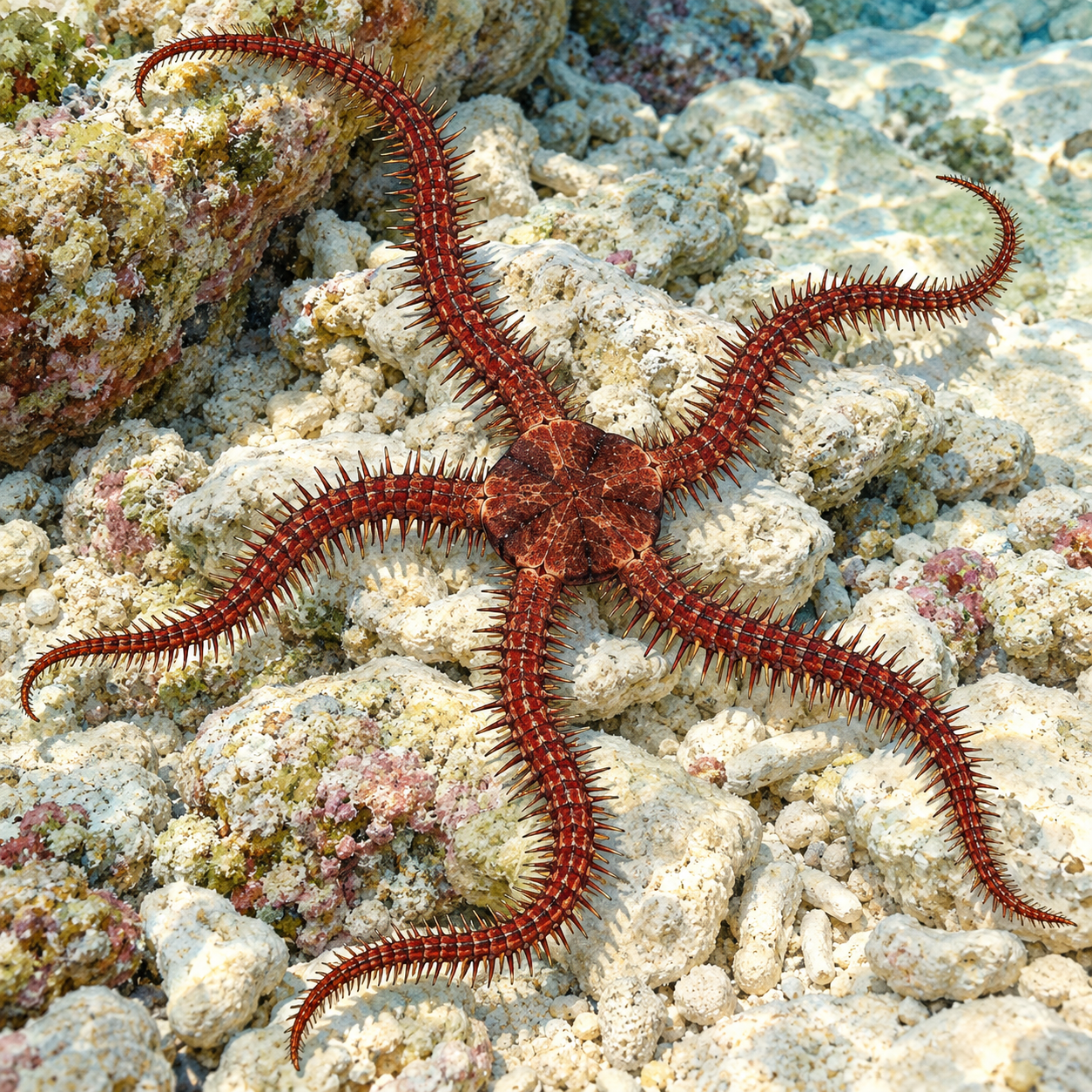
A red-brown brittle star with five spiny arms rests on pale reef rubble.
Natural-history illustration of a brittle star. It is not a photograph of an experimental animal.

A red brittle star moves through a reef with five slender arms, yet it has no familiar pair of eyes. That does not mean it is blind to every useful feature of its surroundings. Research on a Caribbean species has shown that light-sensitive cells spread across the body can contribute to spatial vision.

The distinction is important. Detecting that light has become brighter is not the same as distinguishing where something is. Researchers had to test the animal's behavior to discover whether its distributed sensors could provide information about visual patterns.

From light detection to spatial information

The species discussed in the research was called Ophiocoma wendtii; more recent work uses Ophiomastix wendtii. It changes color between a dark reddish-brown daytime appearance and a paler state at night. Its body is covered with numerous putative photoreceptors.

In experiments reported in 2020, the animals responded to visual stimuli in ways that supported spatial vision. Their responses were coarser than the detailed images humans resolve. A distributed visual system does not imply that the animal sees a sharp, human-like picture of the reef.

For an animal searching for shelter, a coarse distinction can still be useful. Locating a dark area or a suitable refuge may matter more than recognizing intricate details. What counts as useful vision depends on the behavior it serves.

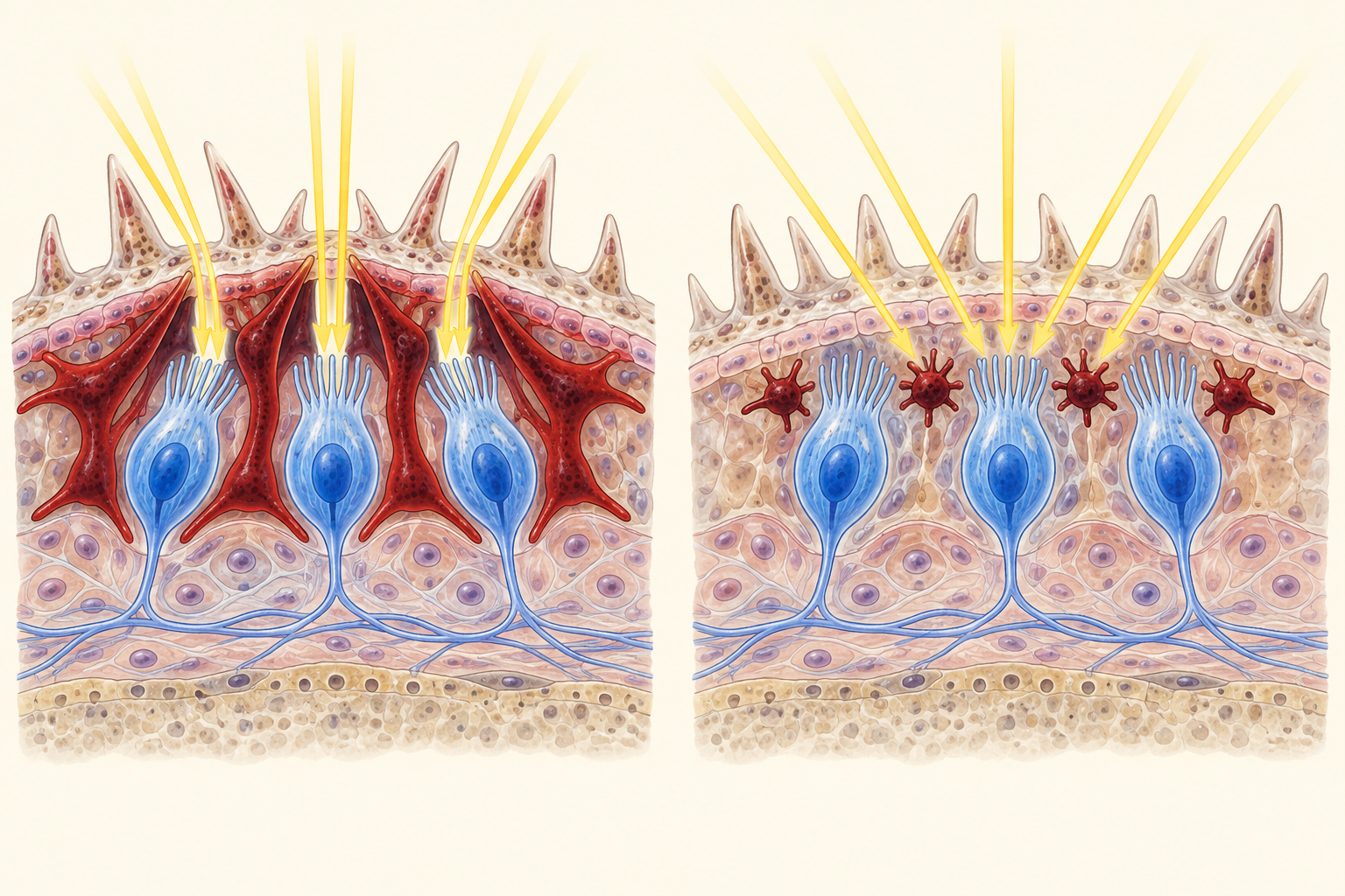
A simplified tissue comparison shows expanded versus contracted pigment around light-sensitive cells.
Conceptual illustration of pigment screening. Expanded pigment limits incoming directions on the left; contracted pigment allows a broader field on the right. Cell shapes and arrangement are schematic.

Color changes the view

The researchers connected visual performance with pigment cells called chromatophores. When pigment is expanded, it can screen light-sensitive cells from some directions. This restricts their fields of view and helps directional information emerge from the distributed network.

In the pale state, contracted pigment provides less screening. The studied animals lost the demonstrated spatial response under those conditions, even though being unable to perform that visual task does not mean every ability to sense light vanished.

A light-sensitive cell needs a direction as well as a signal

Imagine placing one light meter in an open room. Its reading can tell you that illumination changed, but does not necessarily identify which wall became brighter. Restricting the directions from which light reaches a sensor can add spatial information. A group of sensors with different restricted views can then supply relationships that an unrestricted brightness reading lacks.

The pigment-screening account uses this distinction. Expanded pigment limits some incoming directions around receptors. Contracted pigment allows a broader range. No miniature lens is required for that particular change in directional information, although the animal must still use the distributed signals to produce useful behavior.

The diagram isolates that optical relationship. It does not show a measured array of identical cameras, and the illustrated arrows do not establish the animal's visual resolution. Behavioral tests are needed to ask what distinctions it can actually make. Anatomy provides possible machinery; performance tells us what the living animal can do with it.

This helps explain the difference between failing a spatial task and losing every light response. A sensor can remain light-sensitive while the directional arrangement needed for the task is ineffective. The nighttime results therefore sharpened the question about pigment rather than simply proving that the animal had switched all its receptors off.

Why the crystal-eye story changed

Older explanations proposed that small calcite structures in the skeleton acted as microlenses connected to underlying nerves. The idea became a striking example of an animal using its body as an eye. Later anatomical research found that the photoreceptor network was independent of those supposed lenses and did not support their proposed optical role.

Follow-up work continues to examine what this form of vision can achieve and how it functions in the animal's environment. The fascinating claim is already substantial without calling every skeletal crystal an eyeball: an animal lacking discrete eyes can combine distributed receptors, pigment and behavior to obtain useful information about space.

Compare the predictions before admiring the explanation

The crystal-lens story and the pigment-screening story suggest different anatomical relationships. If a skeletal structure were an image-forming lens, relevant light-sensitive elements would need an arrangement that made optical use of it. Later anatomical work found a distributed receptor network that did not support the proposed lens connection. An attractive shape in the skeleton was not enough.

The behavioral comparison adds another level. Dark and pale states, related species and visual targets let researchers ask when the demonstrated response appears. Oxford's account describes how the team considered alternative explanations for the day–night difference, including motivation and illumination. The pigment interpretation gained value by fitting observations across those comparisons.

Read one figure from the linked papers with a specific question: does it show the tissue, the predicted directions of light, or the animal's response to a target? Keep those categories separate on a first pass, then follow the proposed connection between them. It is easier to assess a mechanism when a behavioral result is not mistaken for a photograph of the optical process itself.

The remaining interest concerns what coarse spatial information does for a moving brittle star. A refuge need not be represented in fine detail to be useful. The appropriate comparison is with the animal's actual navigation problem, rather than with the sharpness of a human view or the quality of a camera photograph.

Sources and further reading: Oxford Museum: Color-changing brittle stars can see · Whole-body photoreceptor networks independent of lenses · Extraocular vision and chromatophore movement: original paper · Follow-up: Run and hide, visual performance

Chromatophores: The Muscular Pixels in Cephalopod Skin

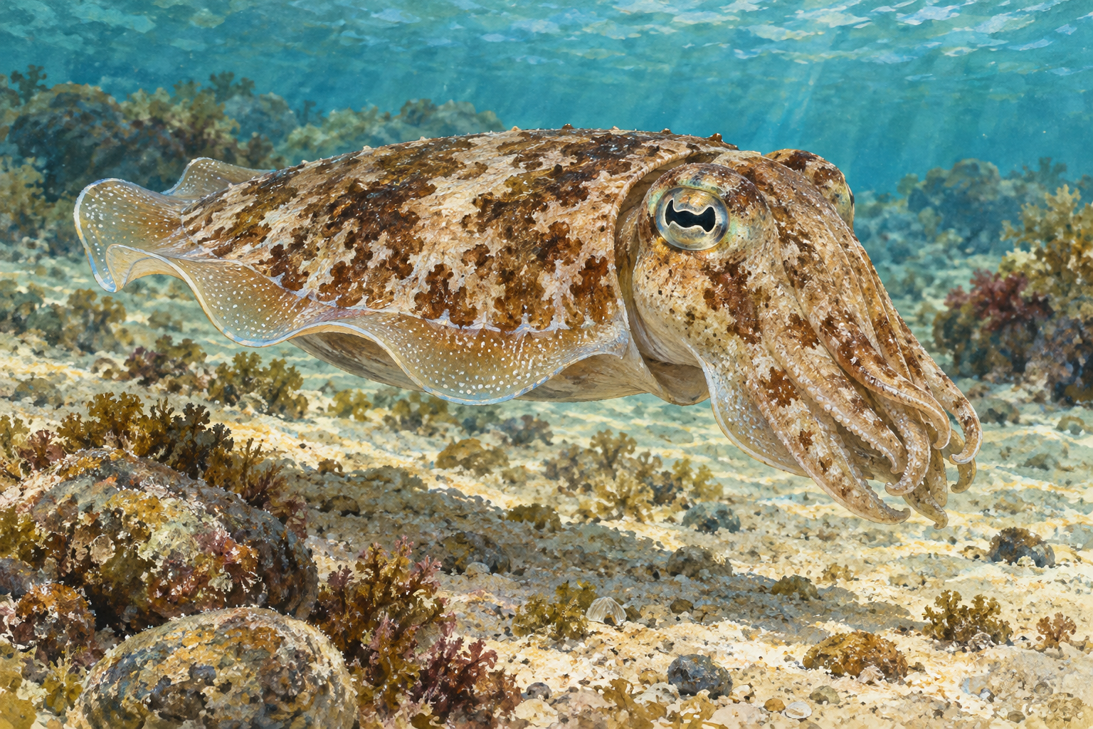
A mottled brown and cream cuttlefish hovers above a stony sandy seabed.
Natural-history illustration of cuttlefish camouflage, not a photograph or a recorded sequence of color change.

A cuttlefish can shift from a pale mottled appearance to strong dark markings while it hovers above the seabed. The transformation can look like an animation playing across its body. A central part of the effect is a set of skin organs called chromatophores.

Calling them pixels is a helpful analogy, provided we remember that each is living tissue. Muscles, nerves, elastic material and pigment work together to change how much colored surface is exposed.

A tiny sac that spreads into a disk

A cephalopod chromatophore contains an elastic pigment sac connected to muscles arranged radially around it. When those muscles contract, they pull the sac outward, spreading it into a broad colored disk. When the muscles relax, the sac retracts and its visible colored area becomes much smaller.

The pigment does not have to be manufactured afresh for every quick change. Mechanical expansion and contraction can expose or hide existing color. Coordinating many such organs creates patches, bands and complex body patterns.

These muscles are controlled through the nervous system. A cephalopod can therefore alter its appearance rapidly as it responds to its surroundings or other animals. The pattern is part of behavior, rather than simply a passive change in the chemistry of its skin.

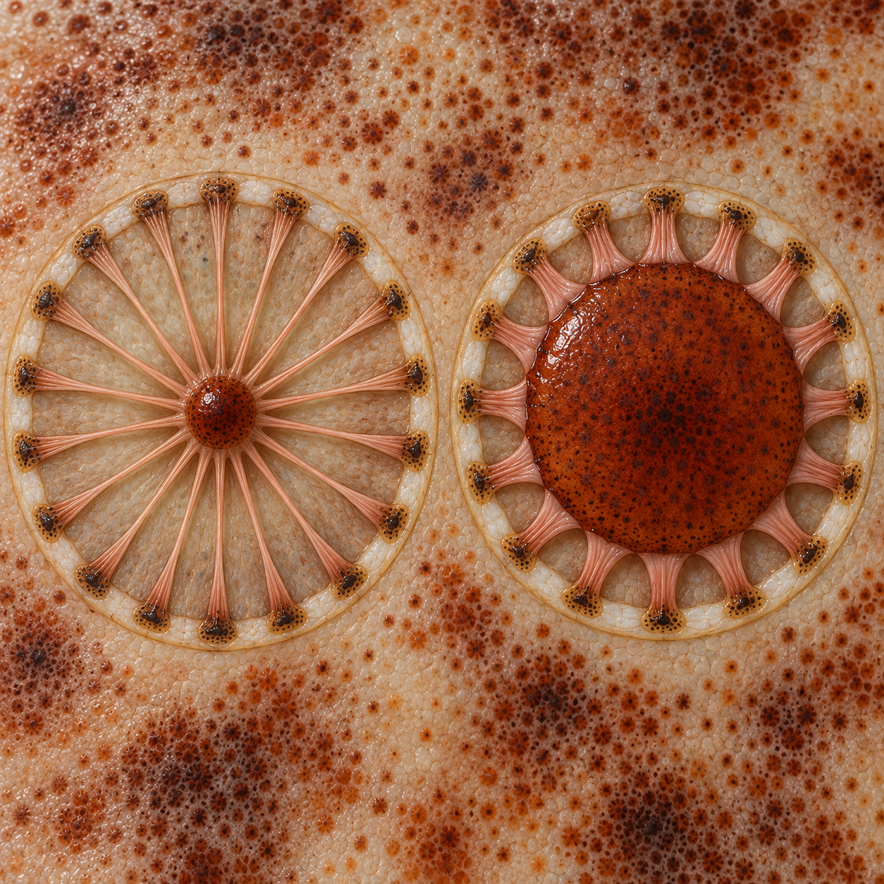
Two conceptual pigment sacs compare a small retracted state with a broad disk pulled outward by shorter radial muscles.
Conceptual illustration of cephalopod chromatophore movement. Left: relaxed muscles and a small pigment sac. Right: contracted radial muscles spread the sac. Anatomy and scale are simplified.

Expanding an existing color changes the visible pattern

A useful way to read the sac diagram is to choose one pigment organ and follow its edge. The colored area expands when the surrounding radial muscles shorten. This can seem backward if we imagine that muscle contraction must always make the visible feature smaller. Here, the muscles pull outward from different directions, spreading the central elastic structure.

The resulting disk changes how much pigment is exposed over a patch of skin. A small dark point can become a much broader dark region without a new supply of pigment arriving at that moment. Coordinated over many organs, changes in exposed area produce a body pattern that can transform much faster than a new coat of pigment could be built from scratch.

The “pixel” comparison captures spatial control, but a screen pixel is not a complete model. The animal's skin has layers and mechanical depth, and reflection changes with illumination and viewing angle. A similar color in two frames can arise through different combinations of pigment exposure and optical effects.

This is why the microscopy is especially useful. It lets us follow the same small structures across time instead of assigning one isolated image to one mechanism. Their changing outlines show a mechanical process, while the accompanying sparkle raises questions about light reflection.

More than one way to make color

Chromatophores are important, but they are not the whole optical system. Cephalopod skin also includes reflective structures. Iridophores produce iridescent effects through microscopic organization, while leucophores can contribute pale or white reflection. The combination gives the animal more possibilities than a layer of pigment alone.

The division is not always as tidy as an introductory diagram suggests. A 2019 study of squid chromatophore organs reported structural coloration associated with the organs themselves, including reflectin proteins in cells around pigment sacs. The researchers found that pigmentary and reflective effects could be intertwined.

This matters when interpreting a shimmering blue or changing sheen. It may depend on how light is reflected, including the viewing angle, rather than on a blue pigment expanding in exactly the same way as a brown one. Species and tissues differ, so a result from one squid should not automatically be treated as the precise mechanism of every octopus display.

Watch the outline first, then the sheen

Live squid mantle filmed by Stephen Senft, Marine Biological Laboratory. Reflective chromatophores sparkle as the incident-light angle changes. Open the research account and both microscopy films.

On the first viewing, choose a dark or yellowish patch and watch its boundary. Does its area change, or does its brightness change while the outline stays broadly similar? Do neighboring patches move together? You are looking for changes that a simple statement such as “the squid turned blue” would hide.

On the second viewing, follow the reflected glints. Notice how their appearance relates to the lighting and the changing tissue. The film is not a claim that every shimmer is produced by a separate blue pigment sac. The 2019 study examined structural coloration associated with chromatophore organs, complicating a tidy division between pigment above and reflectors below.

Finally, compare the microscope view with the cuttlefish scene. The larger animal's outline, posture and skin texture contribute to what a viewer or predator sees. The close view explains a component; it does not reproduce the whole behavior. A cephalopod's remarkable display emerges when these scales are put together, with each observation kept attached to the species and tissue actually studied.

Camouflage is a whole-body performance

Color and pattern can help an animal blend with its setting, interrupt its outline or communicate. Some cephalopods also change posture and skin texture. Cuttlefish papillae, for example, are muscular structures that can make the surface appear more three-dimensional. They are distinct from the pigment organs.

For a second microscopy view, MBL's research page includes microscopy videos of squid skin.

Sources and further reading: MBL: Squid skin coloration strategies · Dynamic pigmentary and structural coloration within chromatophore organs · MBL: How cuttlefish change skin texture

Mudskippers: Fish That Make the Shore Their Workplace

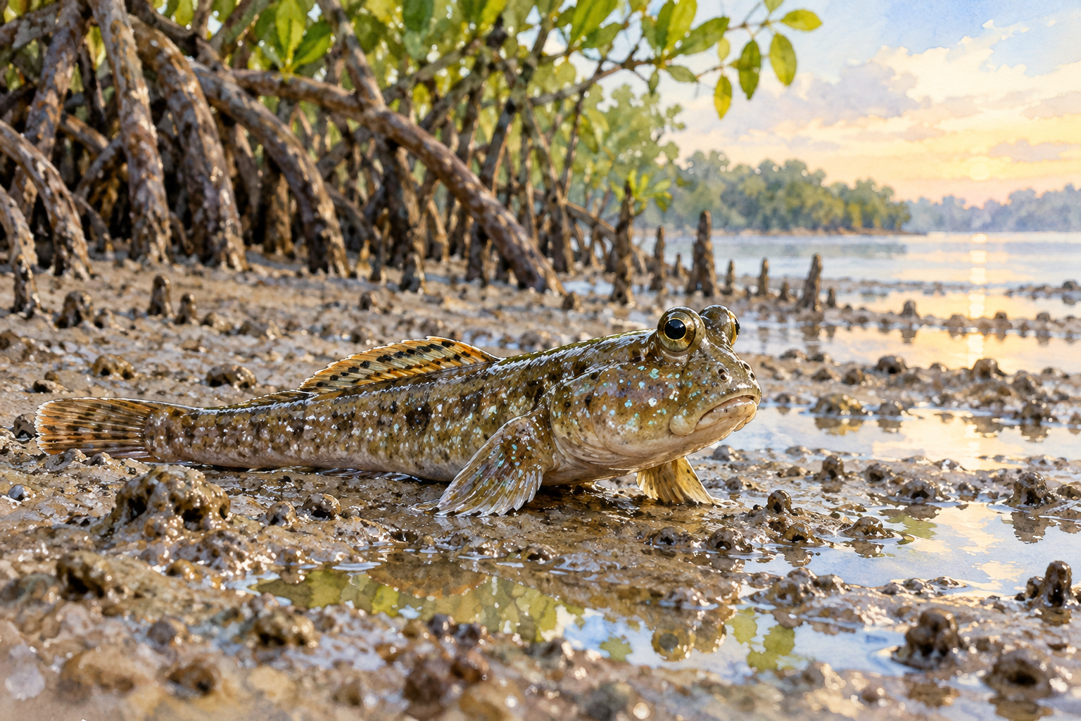
Illustration of a mudskipper resting on its pectoral fins on a wet mangrove mudflat.
A mudskipper-inspired view of life at the water's edge.

Watch a mudskipper for a little longer than seems necessary. At first you notice the obvious surprise: a fish is sitting above the water. Then its paired fins brace against the surface. Its raised eyes move. One eye briefly sinks into its head and comes back out. The more closely you look, the less this seems like a fish making a single daring excursion. It is an animal managing several ordinary problems in an extraordinary setting.

A tidal flat is never permanently one thing. A place exposed to air can later be underwater. For a mudskipper, the boundary offers opportunities, but it also changes how breathing, movement, and exposed tissues work. Understanding the animal becomes more satisfying when we ask what each small action accomplishes.

The shore presents a bundle of problems

Mudskippers are amphibious gobies. “Amphibious” describes their use of both aquatic and exposed environments; it does not place them in the amphibian group that includes frogs. Patricia Wright and Andy Turko’s review of amphibious fishes shows how many different challenges accompany life outside water. Support, hydration, gas exchange, and waste handling all change at the boundary. A solution to one challenge does not automatically solve the rest.

Think first about support. Water supports much of a fish’s weight; on an exposed surface that help disappears. Mudskippers use their fins and body movements to travel over the substrate. They also need moist surfaces for effective gas exchange. The useful question is therefore more precise than “Can this fish breathe air?” We need to ask where oxygen enters, under which conditions, and how the animal keeps the relevant tissues functioning.

These answers vary among species. Research on mudskipper reproduction describes contributions from the gills, the lining of the mouth and branchial cavity, and the skin, in different proportions. There is no single universal mudskipper breathing diagram that explains every species equally well. That variation is part of the interest: related animals can use a shared shoreline lifestyle while differing in how they handle its demands.

A blink built in a different way

The eyes offer an especially good close-up. In a 2023 study, Brett Aiello, Thomas Stewart, and colleagues examined blinking anatomy and behaviour in mudskippers. A mudskipper does not simply close a pair of eyelids like ours. The eye retracts into its socket, where a flexible covering called a dermal cup passes over its surface. Existing eye muscles help produce the movement, alongside this specialized tissue. The original paper connects the origin and function of blinking with life on land.

What would count as evidence that a blink is useful? A neat-looking movement alone would not answer that question. The team tested functions: wetting the eye, removing material, and protecting it. As Penn State’s research account explains, blinking increased when the eyes were dry. Blinks also cleaned the eye and responded to potential injury. The fish lack our tear glands and ducts; water from the surroundings and skin mucus appear to supply a wetting film instead.

That is a valuable distinction between resemblance and mechanism. Both you and the fish can maintain a moist eye by blinking, yet the moving parts and source of the film differ. The resemblance gives researchers a question to test. It does not entitle us to assume that every anatomical detail, or every step of evolutionary history, must match.

Watch the blink, then watch the animal

Watch the research footage linked by Penn State. First follow one eye, rather than the whole fish. Look for its downward movement and the return to a raised position. On a second viewing, widen your attention: is the animal supported above the water, and what can you actually see of the surface around it? Keep the observation separate from the explanation you have just read.

Try making two short notes: “I saw…” and “This could help because…”. The first should contain a visible event. The second is an interpretation informed by research. For example, seeing an eye retract is different from proving that it became wetter. Scientists needed controlled comparisons to investigate that function. A video is a useful invitation to notice, but it does not contain every measurement behind a conclusion.

If your first viewing misses the blink, that is useful information too. A conspicuous evolutionary story can depend on an inconspicuous behaviour. The habit of watching again often reveals more than a new list of surprising facts would.

The nursery with a supply of air

There is another shoreline problem hidden below the mud. A burrow may protect eggs, yet its water can contain very little oxygen. In 1998, Atsushi Ishimatsu and colleagues reported that the giant mudskipper Periophthalmodon schlosseri accumulated air inside its burrows. Their study of stored burrow air identified a way an animal could bring a useful environment into a difficult one.

A later field and laboratory study followed the Japanese mudskipper Periophthalmus modestus. Males added mouthfuls of air to the egg chamber, helping maintain oxygen for developing eggs. When development was complete, the male removed air, allowing the eggs to become submerged and hatch. The authors combined observations with measurements rather than inferring the process from the shape of an empty burrow. Read the egg-incubation and hatching study.

Notice the reversal. Air is useful during incubation; water becomes necessary at hatching. It would be easy to tell this as a story of a fish “escaping water,” but its life depends on controlling when the two environments meet. A burrow can be a refuge, a nursery, and a managed boundary. Findings from a named species deserve that name, rather than being silently turned into a rule for all mudskippers.

A living comparison, not a preserved ancestor

Mudskippers are modern fishes with their own evolutionary history. Their amphibious life and blinking evolved independently of the lineage that produced our four-limbed ancestors. This makes them useful comparisons: researchers can observe how a living animal handles exposed eyes and movement between media. It does not make them a film recording of a transition hundreds of millions of years ago.

The shoreline becomes more interesting when we leave that shortcut behind. A blink, a pause on the mud, and a mouthful of air carried into a burrow are separate actions with separate consequences. Together they show why adapting to a boundary is more than acquiring one spectacular trait. The next time a fish appears to be doing nothing above the waterline, give it another minute. There may be a great deal of work in that small interval.

Find more creative curiosity at Incoprea.

Hagfish Slime: An Emergency Fabric Made in Seawater

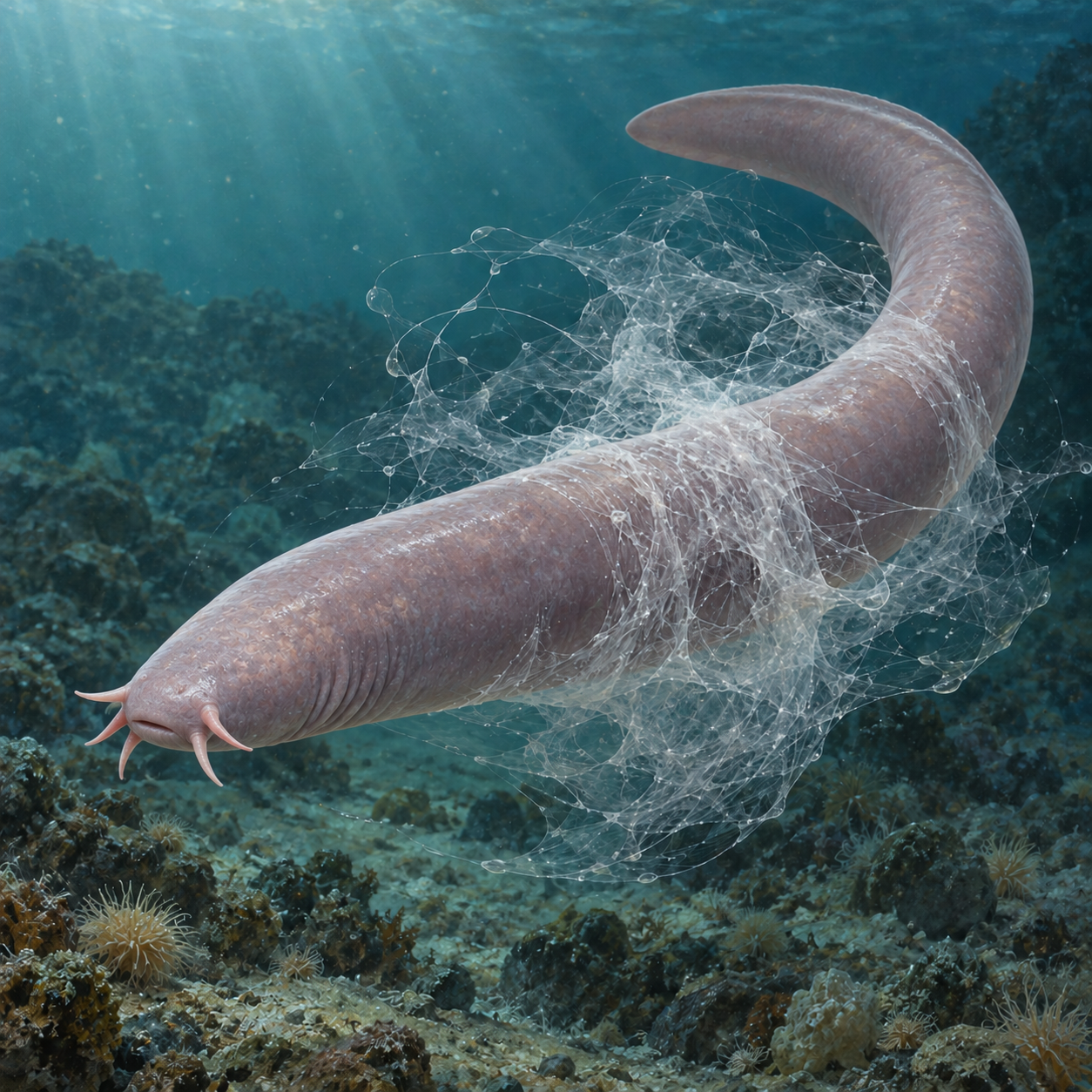
Illustration of a pale hagfish surrounded by a translucent network representing defensive slime.
The fine fiber network is emphasized to explain the material's structure.

A hagfish has a remarkable answer to a predator’s mouth: make the surrounding seawater harder to pump through gills. The animal releases a small amount of concentrated material, and a much larger fibrous slime forms around it. The surprise is not simply the quantity. It is the way a soft, mostly watery structure can briefly interfere with something as urgent as breathing.

Calling this substance “goo” hides the most interesting part. Hagfish slime combines components with different jobs. Some help stop water flowing. Others give the material enough coherence to keep doing that job. To understand it, we need to look at the complete sequence: storage, release, deployment, and the short working life of the network.

A defence tested from two directions

How can we tell that slime actually helps during an attack? Laboratory experiments and observations in the sea offer different pieces of evidence. In a 2006 study, Jeanette Lim and colleagues measured water flow through artificial gill models and real fish gills exposed to slime. The material increased resistance to flow. Their work also showed that mixing mattered: too little prevented proper formation, while excessive mixing could collapse it. Read the gill-clogging experiment.

Field evidence supplies a different view. In 2011, Vincent Zintzen and colleagues reported baited-camera footage of hagfish interacting with other fishes. Their recordings included predators reacting to slime during attempted attacks. The study also documented active predation by hagfish, complicating their familiar image as exclusively passive scavengers. The paper includes the underwater observations.

The distinction is useful. A camera can show that an encounter changes after a release of slime. A controlled flow experiment can isolate a physical effect. Neither should be asked to do the other’s whole job. Together they make the explanation stronger than a dramatic clip or an unusual material test would be alone.

Most of the finished material comes from outside

A hagfish does not store a fully expanded mass inside its body. Specialized glands hold the ingredients that become slime after release. A review by Douglas Fudge, Sarah Schorno, and Shannon Ferraro describes secretory cells producing the mucous component and cells producing protein threads. Seawater becomes part of the resulting material. The threads begin as compact, intricately coiled packages called skeins. Their biomechanics review explains this storage-and-deployment system.

That is a useful engineering idea to turn over in your mind. Imagine packing a tent without carrying the air that will eventually occupy it. The comparison is imperfect: slime does not have poles, canvas, or an enclosed interior. What it captures is a design principle. A compact supply of material can recruit something already present in the environment. The final size need not be limited to the volume carried at the start.

There is another important limit to the analogy. The slime is not simply a superabsorbent powder swelling into a solid jelly. Its components form a network that impedes water movement. “More volume” and “more solid material” are different measurements, and confusing them can turn a fascinating adaptation into an implausible claim about an animal manufacturing matter from nothing.

Mucus and threads do different work

A 2023 study by Luke Taylor and colleagues compared the contributions of the slime’s components. Mucus was central to effective clogging and low permeability, while the threads supplied mechanical strength and helped preserve the clogging function over time. The researchers also compared the slime with other thickening agents. The result was more specific than “very thick slime blocks a hole”: a highly dilute material could still be a strong barrier to flow. Read the component experiments.

Permeability means how readily a fluid can move through a material. Two structures can look equally wet while allowing very different rates of flow. That helps explain why a watery-looking material can matter at a gill. A predator needs a continuing supply of water passing over its respiratory surfaces. Interfering with that flow can be useful even if the defence is temporary and visually insubstantial.

Predict the experiment

Suppose you were comparing a complete slime sample with samples containing its components separately. You would measure two things: how strongly each sample initially resists water flow, and how well that effect lasts under continued disturbance. Would the material best at the first job necessarily be best at the second? Make a prediction before opening the research result below.

Reveal what the component study found

The mucus was especially important for initial clogging and low permeability. The threads helped maintain the material’s mechanical integrity and clogging ability over time. The complete system therefore combines roles. This does not mean either component is useless alone; it means that “works” has to be defined by the measurement and the time span.

Now change your question from “Which ingredient is strongest?” to “Which combination preserves the useful function?” That small change is the point of the exercise. Materials are often judged by one appealing property, even though their real task depends on several properties acting together.

A package must stay closed until it is needed

A coiled thread that unravels inside the gland would be a poor package. In 2019, Gaurav Jain and colleagues tested chemicals found in Pacific hagfish slime glands. In laboratory experiments, trimethylamine N-oxide and betaine inhibited skein unraveling. The results supported the idea that the gland’s chemical environment helps stabilize the stored bundles, possibly through effects on adhesive proteins. The authors presented this as evidence for a mechanism, with appropriate limits on what an isolated experiment proves. Read the skein-stabilization study.

Storage and release pull in opposite directions. Inside the animal, the package needs to remain organized. Outside, it must deploy quickly enough to matter. A successful fibre is therefore only part of a successful defence. The animal also needs a reliable way to keep it compact, then make its hidden length available at the right moment.

What engineers are learning to copy

A 2025 study demonstrated synthetic skeins that unravel under fluid flow. Rather than claiming to reproduce every biochemical detail of hagfish slime, the researchers focused on design principles: coiling, adhesives, deployment in moving fluid, and the dimensions and stiffness needed for a soft network. Their open research record includes supplementary movies that let you compare compact packages with their deployed forms.

A demonstration like this is a meaningful research result. It is also a different achievement from a finished medical, industrial, or consumer product. Manufacturing reliably, controlling deployment, and testing a material in its intended environment would each raise further questions. The biology provides a way to think, rather than a guarantee that every proposed application will work.

The lasting interest of hagfish slime lies in that complete chain of events. It is stored small, released at a critical moment, assembled with help from seawater, and useful for a limited time. Seen this way, a famously unpleasant substance becomes a precise lesson in making the right material only when and where it is needed.

Explore more unexpected ideas at Incoprea.

Famous Last Meals is now in paperback — stories, history and 25 recipes

Famous Last Meals: Stories, History, and Recipes is now available in paperback on Amazon. The illustrated collection brings together 23 his...