A patch of lichen on a branch looks like something with a clear boundary. It may have leafy lobes, a branching tuft or a crust that follows the bark. You can point to it. Yet explaining exactly who lives within that boundary is much harder than recognizing the patch.
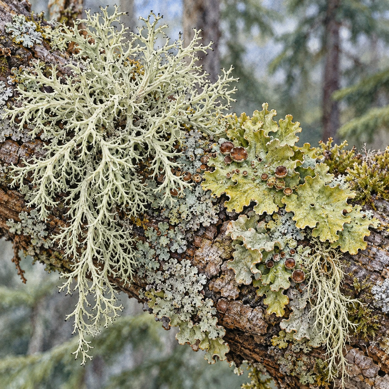
The familiar introduction is a partnership between a fungus and a photosynthetic organism, often a green alga or a cyanobacterium. That introduction remains useful. The fungus and its photosynthetic partners are central to the distinctive lichen body. What has changed is the expectation that the introduction supplies a complete membership list.
The modern research review describes lichen symbioses as open relationships that can include additional fungi and other microorganisms. The scientific task is no longer only to find more names. It is to establish where the organisms are, how consistently they occur and what their presence does.
First find the unexpected resident
A 2016 study identified basidiomycete yeasts in the outer cortex of many sampled macrolichens. These were separate from the familiar ascomycete fungus forming much of the body. Their abundance correlated with differences in lichen appearance and chemistry, and related associations occurred across broad geographical areas.
The location was important. A yeast found in material scraped from a lichen could be a surface arrival or contamination. Microscopic localization inside a particular layer gives the association a more specific biological setting. It changes the question from “was its DNA nearby?” to “what is it doing in this part of the structure?”
A correlation with a chemical difference still leaves several possibilities. The yeast might help produce a compound, stimulate another organism to produce it, benefit from its presence or respond to a shared environmental condition. The discovery opens those alternatives; it does not resolve them simply by being surprising.
Then find a resident without an obvious symptom
In 2019 work on wolf lichens, Letharia, researchers detected an additional basidiomycete group, Tremella. A targeted screen found its DNA in 95 percent of the studied specimens. The work included lichens without the conspicuous galls historically associated with the fungus.
That result makes a quiet-looking surface harder to interpret. An organism previously recognized through a visible growth can also have a much less conspicuous presence. Absence of a gall does not establish absence of the associated fungus. Nor does presence alone settle whether an interaction is beneficial, neutral or costly under all conditions.
Natural history has many such mismatches between what draws our eye and what is common. A dramatic structure is easy to notice, name and collect. A diffuse or microscopic stage needs another method. Our inventory can change when the method stops depending on a conspicuous symptom.
A bigger partnership does not imply one universal third member
A different 2019 survey searched metagenomic data from 413 samples representing 339 lichen species. It detected cystobasidiomycete yeasts in nine species, or 2.7 percent of the sampled species. The authors concluded that these yeasts should not be assumed to occur in every lichen.
The studies differ in their sampled organisms and detection methods. A targeted assay and a broad metagenomic screen need not have the same sensitivity. Those differences deserve investigation, but they do not justify replacing the data with a universal slogan. “More than two partners can occur” and “every lichen requires the same third partner” are very different propositions.
This is a good moment to pause over the word partner. It can mean a regularly associated organism, an organism contributing to a function or an indispensable participant. A careful account says which meaning the evidence supports. The count changes when the question changes.
One common species, a much larger census
A 2025 study of Xanthoria parietina assembled 168 nonredundant microbial genomes across eight lichen samples. The samples came from different substrates and conditions. Some associated lineages were widespread across the samples; others varied with substrate. The number describes the combined recovered catalog, not 168 indispensable organisms inside every individual lichen.
The authors also compared gene expression in the main fungus within lichens and in isolated culture. Expression differed across those settings and among lichen developmental regions. Many functions remain unresolved. Even some detected fungal genomes could reflect propagules on the surface rather than participants building the sampled body.
These distinctions are where the investigation becomes deeper than a new species count. A genome helps identify a possible resident and its capabilities. Gene expression helps reveal which machinery is active. Localization and experiments are still needed to connect an organism with a particular role.
Try a slower look at an ordinary patch
The printable observation sheet below starts with what a hand lens or close photograph can actually reveal. Describe the outline, branching, lobes, discs and substrate. Note whether you are looking at one continuous patch or several neighboring forms. Keep identification tentative when the required features are not visible.
Two scales of lichen observation
A printable sheet separates visible surface descriptions from questions requiring microscopy, sequencing or experiments.
Then turn to the sheet's second column: what cannot be determined from that view? You cannot identify an internal yeast by the lichen's general color. You cannot infer gene expression from a branching silhouette. A beautiful visible surface and a molecular membership list are complementary descriptions, rather than rival ways of seeing.
For a useful comparison, revisit the same accessible patch in different natural light or moisture conditions without removing material. Record what changed in appearance and what remained recognizable. The exercise encourages observation; it is not a diagnostic test for the microbiome or for air quality.
Where does the lichen end?
Researchers also debate how this complexity should affect the definition. A published criticism argues for preserving the distinction between the central lichen-forming partnership and organisms inhabiting its body. A response defends an ecosystem framing as a way to encourage work on the broader set of relationships and physical conditions.
This is more than a dispute over a word, but it need not become a choice between ignoring the central partners and ignoring everybody else. A definition can be useful for naming a biological structure, while an ecological investigation asks who else occupies it. Clearly stating the question lets both descriptions do useful work.
The branch you passed yesterday may therefore contain a familiar outline and an incompletely known community. Its interest does not depend on announcing that old knowledge was worthless. The familiar fungal and photosynthetic partnership is the entrance to a much more crowded, experimentally demanding interior.
Sources and further reading: Evolutionary biology of lichen symbioses (2022); Basidiomycete yeasts in macrolichen cortex (2016); Two basidiomycete fungi in wolf-lichen cortex (2019); Broad metagenomic survey of 339 species (2019); Complexity of Xanthoria parietina symbiosis (2025); The disadvantages of current proposals to redefine lichens (2024); Reflections on lichens as ecosystems (2024).
Explore all 80 new articles · Browse the complete illustrated collection
Explore more art, ideas, and independent projects at Incoprea.